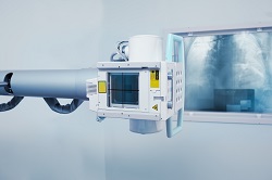
Scaled down X-ray phase-contrast imaging for medical applications and beyond
X-ray imaging has traditionally been working through attenuation – that is, the reduction of the beam as it gets absorbed or deflected by matter. But this has important shortcomings, most importantly the fact that the resulting image lacks contrast. For example, in the medical sector, X-rays have a hard time differentiating between different sorts of soft tissues. ‘By using changes in X-ray phase instead of attenuation, we can increase the contrast of all details in an X-ray image, which in turn allows for the detection of features classically considered as "X-ray invisible”,’ says Professor Alessandro Olivo, coordinator of the LABCAXPCI project. ‘There are many areas where this has key importance: In hospitals, for instance, it would remove the need for the bulkier, slower and more expensive MRI in some applications. It’s a “best of both worlds” situation where one could have soft tissue sensitivity comparable to MRI at the low cost, high speed and high spatial resolution of X-rays.’ LABCAXPCI was all about bringing this powerful X-ray phase-contrast (XPC) technology from over EUR 100 million facilities to standard labs. Where previous attempts at doing so focused on making the source more coherent (which inevitably reduced the emitted flux and resulted in particularly long exposure times to X-rays’ ionising radiations), the project team rather adapted the imaging technology for use with incoherent sources. ‘This allows the use of larger focal spots, automatically leading to higher fluxes,’ Prof Olivo explains. Compared to conventional X-ray methods, LABCAXPCI demonstrates soft tissue contrast – notably the detection of faint tumour features or small damage in cartilage. It enables visualisation of many otherwise invisible features and increases the contrast of visible features by one order of magnitude and above. ‘Compared to other attempts at lab translation of XPC, we typically have shorter exposure times and lower delivered doses, and use imaging systems which are less sensitive to environmental vibrations and more easily scalable to larger fields of view,’ says Prof Olivo. The project’s technology has a wide range of potential applications. In medical imaging, Prof Olivo expects it to eventually become an in-vivo modality for diagnostic and treatment planning, monitoring and assessment purposes. The team is already developing prototypes for the likes of intra-operative specimen imaging which could transform surgical procedures, as well as digital histology. Pre-clinical imaging is also a key application, as many drug development campaigns currently depend on it. Besides medical applications, the Professor of Applied Physics at University College London (UCL) also contemplates non-destructive testing in industry, security scans, or even specialised scientific instruments: ‘We are negotiating with a range of companies to pursue commercialisation in a variety of areas. In medicine, we will initially target specimen imaging (both for intra-operative use and for digital histology), then scale up to in-vivo preclinical and clinical applications. Plans for exploitation in non-destructive testing are also underway, and a first prototype to explore applications in security is being tested.’ A technology licence is currently owned by Nikon Metrology for a variety of areas. Separate licences for in vivo pre-clinical and clinical imaging are currently in advanced stages of negotiation with other companies, with joint pilot studies already successfully completed in some areas
Keywords
LABCAXPCI, X-ray, MRI, soft tissue, synchrotron, XPC, contrast, clinical imaging

