Low-dose 3D X-ray imaging opens new horizons
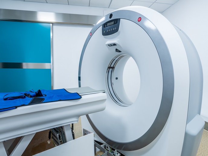
A CT scan uses a rotating X-ray source that captures thousands of views from different angles to construct cross-sectional 3D digital images. This is necessary because the detector only sees the shadow of the whole object for each view.
A novel camera for X-rays
The EU-funded VOXEL project proposed to overcome this limitation by employing plenoptic imaging. As project coordinator Marta Fajardo explains, “our approach records not only the light that reaches the detector, but also from which direction this light is coming. This means that we can compute, with some accuracy, each point of the object, in 3D, just from a single view.″ The VOXEL scientific team are leaders in X-ray metrology, wavefront sensing, atomic physics, mathematical computing and 3D medical imaging. During the project, they developed a novel camera, which consists of an X-ray source, collimating optics to irradiate the object in the most efficient way possible, followed by a main lens, an array to capture the direction of the incoming beam, and a detector. This is the first time this type of camera setup has been developed for X-rays. Researchers had to overcome technical challenges associated with the construction of X-ray optics. They employed engineering and mathematical studies on a prototype using visible light, which helped them constrain the design of the X-ray optics. This led to the generation of two camera prototypes, one for small biological cell microscopy using soft X-ray light, and another for small animal imaging using harder X-rays. Scientists have only just managed to capture the first X-ray plenoptic images from voxels and data processing is ongoing. Nonetheless their measurements have validated the camera design for the retrieval of 3D information at very high resolution using soft X-rays. “Undoubtedly, making plenoptic imaging work with X-rays is our most significant achievement,″ states Fajardo. Additionally, she notes that the scientific teams are very close to making a 3D camera for small-animal or histology applications. A whole human body is not yet possible, and would require further advancements in X-ray optics.
Future prospects of the X-ray camera
According to Fajardo, VOXEL embarked on a very risky adventure four years ago, but the active and tight cooperation of the different teams led to a successful outcome. The acquired expertise and collaborations through the project helped team members establish themselves in the field and also contributed to the training of the next generation of young scientists. By avoiding the rotation of the X-ray source or the sample, the disruptive VOXEL technology enables the use of extremely low irradiation doses for maximum impact on medicine and biology. Despite the prototypes, there are still many design challenges before an X-ray camera is produced for commercial purposes. The VOXEL industrial partner has already secured a future and emerging technologies FET launchpad to study the commercial potential of the X-ray camera. Extra funding will be needed to increase the readiness level of the prototype technology and support its future development from a proof-of-concept to a medical innovation.
Keywords
VOXEL, X-ray, camera, imaging, optic, prototype, plenoptic, dose, irradiation, CT scan