Biological microbe probe
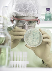
The EU-funded project 'Microbial recognition and adhesion on the nano scale using BIO-SPM' (MICROBIALSPM) has investigated the interaction between live Escherichia coli (E. coli) and its biotic (living) and abiotic (non-living) environment. Scanning probe microscopy (SPM) has put on nanoscale view the physical and biochemical changes experienced by live E. coli. SPM techniques included force spectroscopy, topography and recognition imaging, Kelvin probe force microscopy (KPFM), and scanning microwave microscopy (SMM). The SPM tips were loaded with biologically active molecules to improve specificity at nanometre and pico-Newton resolution. Researchers studied the changes in bacteria and its surface structures when coming into contact with, and colonising biotic and abiotic surfaces in different environmental conditions. Under different growing culture conditions, variations in bacterial morphology and function were studied through changes in surface charge distribution using techniques like KPFM and SMM. The scientists mapped both chemical and mechanical surface properties. Researchers therefore successfully elucidated the underlying mechanisms and the biomolecular interactions involved in these interactions. SPM technologies developed during this project provide unprecedented resolution and sensitivity for imaging live cells or systems in dynamic conditions. Such nanoscale electrical detection methods for biological systems could be exploited for development of miniaturised biosensors and diagnostic devices. Project activities have also laid the groundwork for future studies on disease pathogenesis and drug efficacy. This has significant implications for the material science, surface chemistry, biotechnology and pharmaceutical industries.