High-resolution real-time neuronal imaging
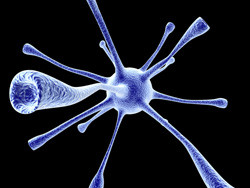
Neurons have a unique morphology compared to most other cells in the body that are an approximate sphere. In addition to the cell body, they have specialised extensions for sending and receiving information. A branched dendritic tree comes off one region of the cell body and a single long axon off another. Cells are small and dendrites even smaller. To complicate the picture a little more, the dendrites themselves have little knobby mushroom-shaped protrusions called dendritic spines. It is here that the synapses or junctions between neurons do their magic. It is also here that many neurological diseases find their origin. Given their extremely tiny size and fast dynamics, studying them in situ has been very difficult. Scientists launched the EU-funded project 'Nanoscale photoactivation and imaging of synaptic spine dynamics' (DYNASPINE) to develop and apply the techniques to do so. Their ultimate goal was to correlate structure and function on the single-synapse level in real time. Neuronal signalling relies on a complicated interaction of chemical and electrical components. Voltages along the membrane change, pores in the membranes open and close, and ions and molecules move in and out. Even the number, size and shape of spines demonstrate plasticity (the ability to change). Such changes can accompany increases in synaptic strength that last for long periods of time (long-term potentiation), also induced by repeated stimulation. This phenomenon is thought to be involved in learning and memory. Scientists applied a combination of electrophysiological recordings and one of the most advanced and high-resolution microscopy techniques available, stimulated emission depletion microscopy. The team uncaged photo-releasable glutamate, an excitatory neurotransmitter, to stimulate receptors at a single synapse. Experiments revealed the plasticity of the spine, in particular shortening and widening of the spine neck, during synaptic potentiation. They also showed that these structural changes had unexpectedly different effects on chemical and electrical signalling, pointing to a new layer of complexity in neuronal dendritic spine function. DYNASPINE opened a new window on functioning dendritic spines. Follow-up of this exciting research direction will be met with great interest by the neuroscience community.