First-time–right structure determination
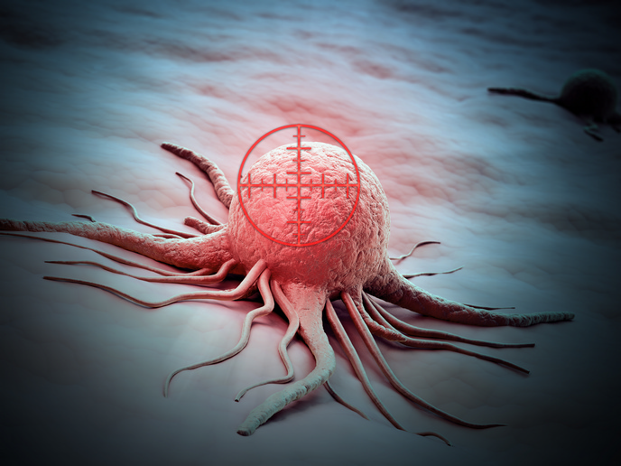
Numerous advanced techniques such as nuclear magnetic resonance (NMR), X-ray crystallography and cryo-electron microscopy (cryo-EM) now make it possible to determine the complex structure of many molecules. However, structural biologists face important limitations inherent in the currently available methods. All three require averaging the results from thousands of molecules, making preparation time consuming and difficult but also rendering it virtually impossible to study rare proteins. X-ray crystallography and cryo-EM cause radiation damage of the samples, and NMR and X-ray crystallography are limited to certain molecular size and shape. The EU-funded study 'Quarternary structure imaging with nano-magnetic resonance imaging (NANO-MRI)' (NANO-MRI) was launched to develop the technology for determining protein structure from a single molecule. Scientists focused on improvements to advanced magnetic resonance force microscopy (MRFM) with nano-scale resolution using the tobacco mosaic virus as the test case. MRFM combines magnetic resonance imaging (MRI) and atomic force microscopy. It is a non-destructive technique with single-particle resolution and the ability to distinguish different elements. It is currently able to reconstruct an element-specific 3D structure. However, its resolution is around 5 nm, making fine details and atomic structure inaccessible. Given that the contrast in MRI arises from differing concentrations of water protons, using two different isotopes of hydrogen is one way to increase atomic resolution. Scientists introduced hydrogen into viruses via the natural route in plants. They synthesised other parts of the virus in bacteria fed with deuterium-containing nutrients to get deuterium into those. The team then successfully produced 'striped' viruses with alternating hydrogen-containing and deuterium-containing parts by discovering the conditions causing the two to self-assemble. NANO-MRI has laid the foundations for non-invasive single-sample structure determination. Achieving this ambitious goal will pioneer a new era of structural biology and structural chemistry, speeding the rate of analysis and revealing structures of molecules previously inaccessible with other technologies.