Spatial distribution of biomolecules in 3D
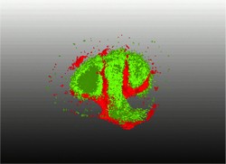
Three-dimensional imaging has a significant impact on many challenges in life sciences. Existing techniques display organ anatomy by tracing the localisation of a specific compound in the body such as water or other labelled molecules. However, these methods are less useful in proteomics or metabolomics studies that are oriented towards identifying new biomarkers, drugs, and disease pathways. Matrix-assisted laser desorption/ionisation imaging mass spectrometry (MALDI-IMS), is an emerging imaging technology that can detect the spatial localisation of molecular ions. 3D MALDI-IMS is a label-free method that can complement other techniques for spatial identification of molecular compounds. However, the analysis of the large and complex data it generates requires new statistical methods. Towards this goal, scientists on the EU-funded 3D-MASSOMICS(opens in new window) (Statistical methods for 3D imaging mass spectrometry in proteomics and metabolomics) project set out to develop statistical methods for unsupervised and supervised analysis to improve results' reproducibility. These methods were tested using data from diabetes, surgical metabolomics, and microbial metabolomics. The consortium developed a statistical simulator of MALDI-IMS data on the rat brain and validated it with additional data from the three biomedical applications. These provided the much-needed support for methods development, allowed scientists to perform relevant data collection, and carry out expert evaluation. Among the developed scientific methods was an innovative approach for measuring spatial chaos, which was applied for the detection of mass-to-charge images. This method represented the first unsupervised univariate analysis of MS imaging. Researchers used these methods to study in 3D the interaction between the microbes Candida albicans and Pseudomonas aeruginosa — microbes implicated in cystic fibrosis (CF). Their observations led to the detection of a molecule (rhamnolipid) that could be exploited for the therapy of CF. Overall, the study succeeded in their vision to develop statistical methods for analysis and interpretation of 3D MALDI-IMS data. Implementation of these tools should make 3D label-free proteomics and metabolomics feasible and improve disease diagnosis.