A new perspective on visualising heart development
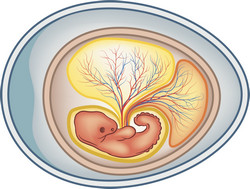
Formation of the vertebrate heart during development is an intricate process involving the complex interaction of genetic and epigenetic factors. From commitment to cardiac cell fate to valve formation, it has to be carefully and infallibly regulated. Mistakes in any step of the process could lead to congenital heart disorders. Avian heart morphogenesis resembles the mammalian process, and the avian and mammalian heart share similar four-chamber architecture. As a result, the avian embryo represents a suitable model for studying heart development and its optical accessibility permits direct observation of the cellular movements. The DYNIMHEART (Defining the biomechanics of the developing heart through high-speed dynamic fluorescent imaging in transgenic quail embryos) project aimed to further advance this system. This was achieved through dynamic fluorescent imaging of the developing vascular system. Scientists created transgenic quails that expressed fluorescent proteins in the endocardium or endothelial cells. By using the spectral signature, it was possible to map their movement and distinguish them from other cell types. Specialised software was developed that facilitated fluorescent cell tracking and together with high-speed confocal microscopy enabled data to be reconstructed into 4D images. Researchers also developed a novel technique called enhanced Number and Brightness (eN&B), which built upon previous (N&B) protocols. The technique enables the aggregation of EphB2 receptor in living cells to be imaged to an unprecedented level of resolution. The technique is not limited to EphB2 and may be used in any protein that can be tagged fluorescently. By implementing it in living embryos, scientists will be able to determine the key players involved in tissue separation and morphogenesis. Particular attention was given to activation of the Ephrin pathway, an essential component of tissue boundary formation. As an indicator for Ephrin activation, the consortium used the phosphorylation of tyrosine kinase receptors. In addition, detection of receptor aggregation in real time provided a further perspective for studying cell-to-cell interactions during tissue morphogenesis. Results from the DYNIMHEART project will provide the scientific community with a novel tool to study morphogenesis, giving unprecedented insights into the spatiotemporal development of the heart. They should also unveil novel molecules with a fundamental role in cardiac morphogenesis.


