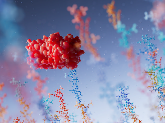
Delineating membrane protein conformation

Membrane proteins serve as channels for the internalisation of ions, nutrients and drugs. In addition, they receive signals from the environment and transduce them to the inside of cells, contributing to intercellular communication. Nearly 50 % of drugs function by binding to membrane proteins demonstrating their medical importance but also highlighting the need for structure determination. However, the hydrophobic nature of membrane proteins is incompatible with established structural analysis techniques such as X-ray crystallography and NMR spectroscopy. The EU-funded MEMBRANE PROTEINS (Visualizing the structure and function of elusive membrane receptor proteins of the human cell) project worked to develop an alternative method for determining the conformation of membrane proteins. In this context, researchers employed a powerful alternative technology based on mass spectrometry to measure the hydrogen/deuterium exchange (HDX) of proteins in solution. Using this technique, the consortium examined the structure and interactions of the T-cell receptor (TCR), which participates in antigen presentation and is pivotal in human immune responses. By estimating the differences between soluble TCR and TCR bound by relevant antigen-loaded MHC complexes, scientists obtained invaluable structural information. Results revealed that both the constant and the variable domains in both chains of the TCR had a highly protected core. Upon MHC complex binding, scientists observed reduced HDX in several distinct regions of the TCR, indicating that these regions participated in the binding. As expected, the complimentary determining regions (CDR) loop structures of the variable TCR domain, known to be responsible for antigen recognition, were protected from HDX. Further experimental evidence showed that each loop contributed to MHC complex binding to a different extent. This would be useful for designing new TCR variants. Overall, the MEMBRANE PROTEINS method provided detailed insight into the structure and function of the TCR, and can extend to other membrane proteins. This could be utilised to design new TCR variants for therapeutic use.