Innovative tools automate brain image analysis
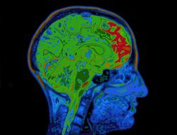
7T MRI provides brain structure definition and tissue composition information with unprecedented high resolution that brings with it its own challenges. For instance, increased data size and stronger field inhomogeneity artefacts can seriously hamper effective analysis. The HIRESBRAIN7T (High resolution segmentation of brain MR images at 7 Teslas) project team have worked to develop suitable automated image processing methods to overcome such issues. During the first project phase, researchers developed segmentation algorithms to process MR images at 0.4 mm. This is impressive, considering the fact that this software handles 15 times more data than the current state-of-the-art. Moreover, they introduced a cortical model of depth that should aid in accurately representing the biomechanics of cortical folding. Using their newly developed tools, HIRESBRAIN7T studied cortical architecture in different areas. They also determined a novel method for alignment of cortical anatomy using intra-cortical contrast. Quantitative T1 with susceptibility mapping created a comprehensive atlas of the tiny but complex basal ganglia. The internal anatomy of its smaller nuclei and segmented other smaller structures such as the hypothalamus or the dentate nucleus were also investigated. Visualising vascularisation is key to understanding brain function as well as detecting pathologies such as cancer. Project members developed new techniques for this purpose. They successfully extracted the venous vasculature from MR images of quantitative susceptibility and modelled the local oxygenation within each vein before it enters the cortex. Most of the HIRESBRAIN7T datasets and tools are freely available online at Openscience(opens in new window) and NITRC. Potential applications include large scale studies of normal ageing, multiple sclerosis, Alzheimer's disease and small vessel cardiovascular disease.