An atlas of foetal brain development
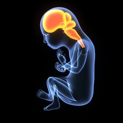
The development of the human central nervous system is a complex process that begins in utero and continues until the end of adolescence. Brain maturation is concomitant with modifications of the cortex and subcortical region during the foetal period. Studies aimed at comprehending brain development are of great clinical interest for the diagnosis and treatment of related diseases. The scope of the EU-funded FBRAIN (Computational anatomy of fetal brain) project was to model brain maturation by integrating spatial and temporal anatomical information into an atlas. In this context, researchers exploited the non-invasive nature of magnetic resonance imaging (MRI) to investigate the developing human brain. MRI complemented ultrasound in brain morphometry and more specifically in studying cortical thickness, myelination and white matter fibre formation. By using new image processing tools, scientists combined the morphological information, as obtained by different MRI images, with diffusion information, to analyse the various anatomical features of the maturing foetal brain. Using mathematical models, they reconstructed high-resolution 3D images and extracted features of brain maturation. By doing so, they successfully estimated all the neural connections of the human foetal brain. Collectively, the FBRAIN tool can help elucidate how brain connections form and evolve in time. Importantly, depicting brain development should help us understand brain pathologies and the emergence of cognition.