Gold trackers for stem cells in a live organism
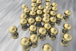
Stem cell based therapy has unparalleled potential in RM for a great variety of diseases. However, a large number of stem cell therapies fail before they can be translated to the clinic. The ability to trace the fate of transplanted cells and monitor grafting efficiency would facilitate clinical translation. The EU-funded NANOSTEMCELLTRACKING (Nanoparticle probes for photoacoustic tracking of stem cell) project developed a new method to track stem cells in vivo using gold nanorods as cell labels and an imaging technique called multispectral optoacoustic tomography (MSOT) for detection. Application of MSOT includes irradiation of the object with pulsed light, whose absorption generates a thermal expansion. This results in pressure waves that ultimately generates an ultrasound signal for transformation into an image. Thus, this technique uses light as excitation source and ultrasound for detection, providing an excellent resolution at depths of up to 5 cm inside the tissue. Researchers used gold nanorods as contrast agents for MSOT because they have a strong absorption in the near infrared, the region of the spectra where biological tissue absorption is minimal. The initial stage of the project involved optimisation of the synthesis of gold nanorods for stem cell labelling. In particular, researchers modified nanorods with silica shells to prevent intracellular aggregation. Toxicity assays showed that modified nanorods were not toxic and did not affect the differentiation potential of the stem cells. In vivo evaluation demonstrated an extraordinary sensitivity of the whole cell labelling and detection system. It was able to monitor as few as 10 000 cells labelled with gold nanorods for 20 days with a resolution of 200 micrometres. In contrast to other optical-based techniques, MSOT enables tracking of labelled engraftments with an excellent 3D spatial resolution deep inside the tissues. Additionally, researchers evaluated the potential of gold nanorods for simultaneous labelling and detection of several cell populations. They used nanorods with a different aspect ratio to label different cells and monitored them in vivo. The preservation of the absorption characteristics of the nanorods allowed to distinguish the two cell populations and to perform co-localisation assays. The project findings are important for areas of experimental and clinical oncology, which can benefit from the ability to monitor cells. The possibility to track cells in real time might help to better understand their immunotherapeutic potential as well as optimise the design of drugs.