Applying cutting edge imaging to real life challenges
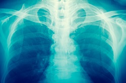
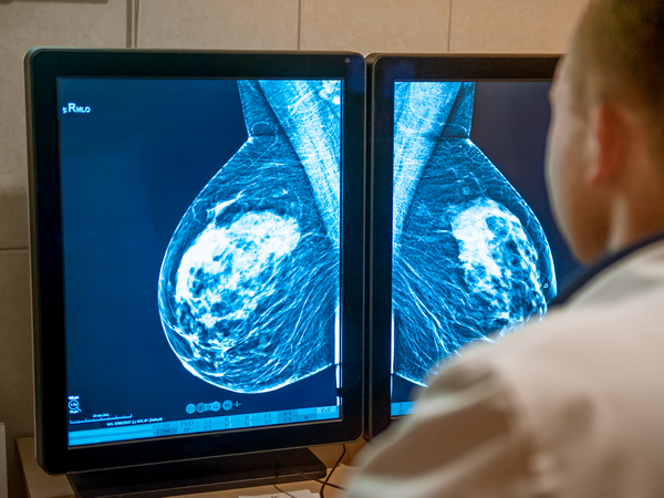
The EU-funded MAXPCI project successfully demonstrated that a technique called x-ray phase-contrast imaging (XPCI) can significantly increase contrast in an x-ray image. This means that features classically considered "x-ray invisible" can be detected. ‘All areas where x-ray imaging is used stand to benefit,’ says project coordinator Prof Sandro Olivo from University College London (UCL) in the UK. ‘People are familiar with medicine and airport security but there are so many other fields – biology, research, cultural heritage, materials science – you name it.’ A range of applications The key success of the project has been to demonstrate that the XPCI technique developed by the team can yield very high phase sensitivity and high resolution, yet at the same time be adaptable to conventional systems (and thus be implementable across a range of fields). This is significant as historically the method has been restricted to specialised facilities. ‘In the initial phase of the project, we were able to demonstrate high phase sensitivity,’ says Olivo. ‘Later in the project this was put into practice in order to demonstrate the potential of this technique in real world applications. One of our most important results was to show that mammography doses could be reduced by more than one order of magnitude.’ The project also demonstrated that an optimised application of the XPCI method can lead to lab-based phase micro CT (computerised tomography) scans to be performed in just a few minutes. CT scans involve a series of X-ray images taken from different angles to create cross-sectional images. ‘All previous attempts at this have taken several hours,’ says Olivo. ‘This is a key breakthrough as it makes the technology compatible with pre-clinical imaging for, say, small animals, and for intra-operative specimen imaging, which can reduce the rate of required re-operations in e.g. breast conservation surgery’. Ultimately the team’s aim is to target in vivo clinical imaging, which will help to improve diagnoses, treatments and assessments in healthcare. Other potential real world applications were investigated during the project. ‘The potential impact is huge and goes well beyond healthcare,’ says Olivo. ‘Pre-clinical imaging can be used in the development of new drugs and the improvement of existing ones, and use of XPCI in security inspections could reduce threats at airports and other sensitive public spaces.’ The technique could also be use in non-destructive testing (NDT), which would enable industry and researchers to evaluate the properties of materials, components or systems without causing damage, and thus save money. Scanning the future The positive results of the MAXPCI project, which was completed in May 2017, will continue to be built upon as Olivo and his team look to make XPCI implementable across a wide range of sectors and industries. ‘We are indeed working with a number of major companies at present to pursue commercial translation in various areas,’ he says. ‘Pre-commercial prototypes already exist and more are under development, which will target many of the applications already discussed. It is possible that other opportunities might arise in the meantime.’ As well as licensing out the technology to various companies to target broader applications, Olivo and his team are also currently considering university spin-outs in specific niche areas. ‘In parallel with this, we are continuing to both improve our approaches and develop new ones in order to target increasingly complex imaging problems. This will enable new research questions to be answered and may provide the basis for future technology transfer.’