Mechanistic insight into cognition in mammals
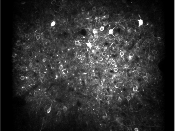
The largest and most complex region of the mammalian brain, the cerebral cortex folds into a big surface of grey matter which comprises neuronal cell bodies organised into a complex circuit network. The anatomical organisation of cortical areas corresponds to its sensory, motor or associative functions. Communication with other brain structures such as the thalamus and basal ganglia ensures the reception and transmission of information. Cortical neurons do not operate alone, but as part of several interconnected assemblies, the activity of which is only partly controlled by sensory stimuli. Understanding the structure of their activity requires the simultaneous recording of many neurons. However, the precise nature of the neuronal interconnections within the cerebral cortex remains elusive. Currently, a number of technological advances are employed to explore cortical computation including electrophysiology, the use of multiple electrodes to record groups of neurons, and optogenetics. The latter method uses light to control neuronal activity in an experimental setting where neurons are genetically modified to express light-sensitive ion channels. However, existing optogenetic tools lack layer and cell-type specificity, while the electrophysiology recording techniques still do not attain the yields necessary to properly characterise the cortical microcircuit. Advanced tools aid understanding of cerebral function The EU-funded NeuroSeeker(opens in new window) project was designed to portray a more functional picture of cerebral circuits. Researchers investigated the structure and function of the cerebral cortex to better understand the cortical areas and the basic laws of multi-scale interactions between them. The consortium wished to describe the interaction of cortical micro-circuits with the global state of the brain by combining recording data with large-scale pictures of the brain state obtained by electrocorticography or electroencephalography (EEG). Towards this goal, the researchers developed ultra-high density, silicon-based probes with an unprecedented amount of ca. 1400 recording channels for electrophysiological recording. Additional polymer-based probes for stereo EEG were generated similar to those that are clinically used in epilepsy diagnostics. The NeuroSeeker probes were used as advanced recording devices with a ten-fold higher electrode count compared to the state of the art. These devices were used in non-human primates to demonstrate recording durations similar to those required for human applications. Furthermore, optical devices were generated comprising integrated thin-film light-emitting diodes (LED) as well as conventional LED chips. These devices produced a controlled optical stimulus to cortical tissues in rodent experiments. ‘To obtain a mechanistic understanding of fundamental computations underlying cognitive processing, we concentrated on candidate mechanisms that map onto common cognitive processes.’ explains Dr Ruther. In brief, researchers investigated the combination of feedforward and feedback inputs that integrate across cortical layers, or that arrive in the same layer from different cortical areas. This allowed them to formulate a dynamic theory of micro-circuits and their interactions with the rest of the brain. Delineation of cerebral cortex Through specific algorithms and software, NeuroSeeker devices enabled the detection and sorting of spike waveforms towards efficient circuit analysis. Contrary to existing systems, the consortium succeeded in recording neuronal function, providing a more detailed view of brain functionality and also advancing epilepsy diagnostics. Investigation of the spatiotemporal fingerprint of each neuronal signal allowed signal classification as well as characterisation and localisation of cells in cortical layers. This provided fundamental information on cortical function in various contexts, from memory formation, to ongoing processing during decision making. Efforts are ongoing to ensure commercialisation of the NeuroSeeker probes through the SME partner. Researchers are hopeful that ‘the NeuroSeeker optical tools might be used not only in animal research but ultimately be applied clinically on the heart muscle as well as in the cochlea.’