

Loosening up tumour cells could stop their proliferation
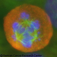
Cancer cells are very good at proliferating, but they're not very good at dividing neatly into two new cells. A team of researchers at the German Cancer Research Center in Heidelberg has investigated this weakness in depth. They found that the tension of the protein fibres involved in cell division is a key feature that allows cancer cells to thrive. The results were published in the journal Science Translational Medicine. During cell division, a normal cell divides neatly along one axis, on a bipolar scaffolding structure. For the division to go on correctly, the cell has two copies of an organising structure called the centrosome. Protein fibres form from the two centrosomes, and pull each of the two sets of chromosomes to one pole. Seen under the microscope, these fibres have the shape of a spindle. Cancer cells, however, often have more than two centrosomes, so the spindle fibres cannot organise properly and take a multipolar shape instead. Such malformed spindles distribute the chromosomes unevenly among the daughter cells, which cannot survive. Some cancer cells have found a way around this: they group several spindles together into two clusters so the cells can go on to divide properly and become thriving tumours. Professor Alwin Krämer, head of a Clinical Cooperation Unit at the German Cancer Research Center and Heidelberg University Hospitals, believes this trick is an underrated Achilles' heel of cancer cells. His team investigated this weakness in detail to identify the proteins necessary for cancer cells to form such clusters. These proteins could be targets for new drugs - if the cells can't form clusters with their extra centrosomes anymore, they'll divide anarchically and die. The scientists screened all the genes of a carcinoma cell line; they switched off each of the 21 000 genes 1 by 1, and looked under the microscope to determine which cells had more than the usual 2 spindle poles during cell division. They identified a series of 82 genes involved in the formation of extra centrosomes. Many of them are involved in attaching chromosomes to the cell's scaffold. In particular, the team believes that the spindle's tension is a necessary feature to bundle centrosomes. Only tightly stretched spindle fibres will place centrosomes close enough to each other for clusters to form. A whole range of proteins are responsible for this tension; if their genes are switched off, the fibres' tension disappears, and cancer cells cannot divide properly. Scientists have known about cancer cells' extra centrosomes for about a century now, but there remain many questions about how this affects cell division and the survival of tumour cells. These findings suggest that some existing cancer drugs are efficient precisely because they interfere with spindle tension in tumour cells with extra centrosomes. Further down the line, this research could help develop more targeted cancer drugs. 'Such a therapy would hit the cancer very specifically, because only tumour cells have extra centrosomes and depend on the survival trick of clustering,' explained study leader and co-author Alwin Krämer from the German Cancer Research Center.
Countries
Germany