Fluorescent viral tracking uncovers yellow fever and Zika virus infectivity
Flavivirus infection starts when the viral particle is internalised into cellular vesicles known as endosomes. After fusion of the viral membrane, the viral genome is released into the cytoplasm of the cell and virus replication begins. This point marks the detection of infection and initiates the cellular response to the virus. However, the precise in vivo dynamics and interactions with the host remain unclear and require quantitative and high-resolution imaging methods.
Super resolution microscopy unveils viral entry into the cell
With the support of the Marie Skłodowska-Curie programme, the FLAVIC DYNAMICS project developed a methodology of labelling viral components to track cellular infection by flaviviruses that include West Nile, dengue, Zika and yellow fever virus. “Using the yellow fever virus as a model, our goal was to dissect the infection process and delineate the role of the different cellular and viral components,″ explains project coordinator Giovanna Barba Spaeth. Post-doctoral research fellow Joao Freire used a combination of fluorescent dyes to label the surface of the virus, lipophilic fluorescent dyes to label the viral membrane and RNA-specific dyes to mark the viral genome. In collaboration with Christophe Zimmer, he was able to visualise flaviviruses through super resolution techniques such as photo-activated localisation microscopy, which can achieve a resolution up to 20 nm. The scientists tracked single viral particles to monitor the viral entry kinetics into host cells. Comparison of the entry kinetics between the wild-type yellow fever and the attenuated vaccine strain 17D, indicated that the mutation in the major virus envelope protein E causes the two viruses to fuse in different endocytic compartments. This means that their presence is detected by the cell in a different way with potential consequences in the downstream stimulation of immune responses. With respect to the route of infection of Zika virus, the researchers, in collaboration with Chiara Zurzolo, identified an alternative cell-to-cell spread path through tunneling nanotubes, cell protrusions that connect adjacent cells.
Clinical significance of FLAVIC DYNAMICS
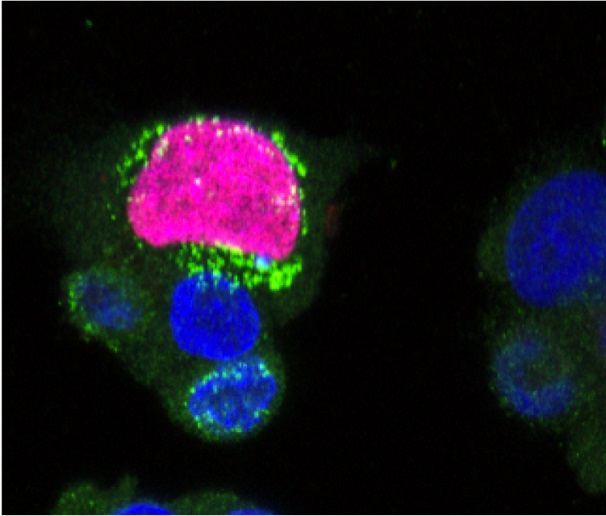
The findings of the FLAVIC DYNAMICS project provide novel insight into the molecular mechanisms leading to yellow fever vaccine immunogenicity. These results may offer a common platform for the attenuation of other pathogenic flaviviruses for which a vaccine is not available. Zika virus attracted mass media attention in 2016 due to its association with microcephaly in newborns. The precise mechanism through which the virus is vertically transmitted between pregnant women and their foetuses is unclear. The discovery that the virus can be transmitted via cell-to-cell contact between different cell types and tissues constitutes a major breakthrough. It provides clues about viral tropism and pathogenicity and may increase its infectivity toward non-permissive cells. “It also suggests that this transmission path may protect the virus from the action of circulating neutralising antibodies,″ emphasises Barba-Spaeth. This has profound consequences for antiviral therapy as strategies designed to block circulating virus must be reconsidered. Future plans include investigation into the structural determinants that cause the different infection kinetics of wild type and vaccine yellow fever viruses. This approach is expected to generate a blueprint for flavivirus vaccines and drive research towards strategies for viral clearance. In the image above, human cells are red and blue and are infected by Zika virus, the green colour.