Novel approach improves resolution in ultrasound images
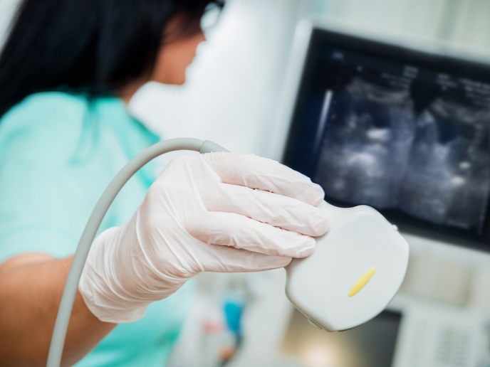
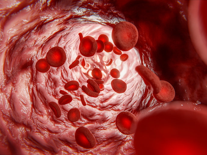
Ultrasound imaging uses the reflection of waves to produce images of soft tissues within the body. However, inherent variations in soft tissue structure can distort conventional ultrasound wave fronts, leading to inevitable blur in the images that can be detrimental to medical diagnosis. With the support of the EU-funded projects REMINISCENCE and SMART, a team of researchers has developed a new non-invasive ultrasound method that overcomes this problem of image quality degradation. The researchers have deployed a universal matrix approach for wave imaging, controlled by a multi-sensor network. The details of their technique were published in the journal ‘Physical Review X’ (PRX)(opens in new window). The results of the study were also published in the journal ‘Proceedings of the National Academy of Sciences’ (PNAS)(opens in new window). Referring to the PNAS article, a press release(opens in new window) by REMINISCENCE project host and SMART project coordinator the French National Centre for Scientific Research (CNRS) notes that “the scientists showed how this new method can subtly compensate for the distortions that a focused wave undergoes as it travels through the studied tissue, with an ideal resolution and contrast optimized for each pixel in the image.”
Various applications
The CNRS press release states: “Applications range from biomedical diagnosis to optical microscopy, detection of cracks in industrial materials, and the monitoring of volcanoes and fault zones in geophysics.” In the PRX journal article, the researchers emphasise that the potential of their work goes beyond ultrasound imaging. “The experimental proof of concept is performed with ultrasonic waves, but this approach can be applied to any field of wave physics for which multielement technology is available. The concept is that focused beam forming enables the synthesis, in transmit and receive, of an array of virtual transducers which map the entire medium to be imaged.” The scientists conclude that their matrix approach “could be applied to any kind of system for which emission and detection of waves can be varied in a controllable way.” They believe the new method has “immediate foreseeable impacts in a range of wave physics including optical microscopy, radar, and seismology.” The ongoing REMINISCENCE (REflection Matrix ImagiNg In wave SCiENCE) project that supported the study will end in May 2024. It was launched with the ultimate objective of developing “an information theory of wave imaging,” as explained in the project factsheet(opens in new window). In addition to “direct imaging applications, this project will also provide a high-resolution tomography of the wave velocity and a promising characterization tool based on multiple scattering quantification.” The SMART (Scattering Matrix Approach in Reflection applied to Turbid media) project ran between 2017 and 2019. It focused on “multiple scattering and distortion of classical waves, with the goal of improving imaging and characterization of complex biological materials,” as noted on CORDIS(opens in new window). Project partners believe that the approaches to imaging used in SMART have contributed to the improvement of biological imaging in areas like imaging of the human eye and skin. “Notably, we have introduced novel imaging modalities, consisting of a focusing criteria enabling refractive index tomography, and multiple scattering quantification.” For more information, please see: REMINISCENCE project(opens in new window) SMART project(opens in new window)