A perfect cage-like nanostructure could ship drug molecules into cells
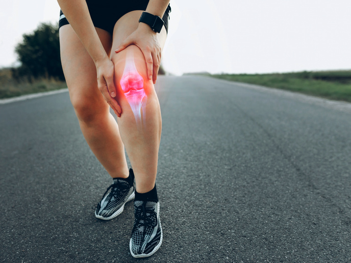
Nanotechnology offers a promising way to deliver and control the release of new drugs and a tremendous opportunity to overhaul old treatments by improving their safety and efficacy. Functional nanoparticles encapsulating drug molecules can selectively reveal their therapeutic action close to the target tissue or cell without flooding the bloodstream with medicine.
Helping nanoparticles deliver on their promise
The aim of the NanoIntra project, which received funding under the Marie Skłodowska-Curie Actions programme, was to clear a key hurdle to making nanoparticle drug delivery a reality. Besides achieving a fine control and release of therapeutics on the nano level more accurately, we need to understand where the drug is trafficked once the nanoparticle is taken up inside the cell. In particular, drug molecules can be rendered completely ineffective if they remain in the endosomes(opens in new window). Merging expertise from different fields, such as chemistry, biology and physics, NanoIntra scientists created several innovative nanoparticles including luminescent semiconductor quantum dots, plasmonic metal nanocrystals and silica nanoparticles for accurate bioimaging and biosensing analysis. In a second step, they functionalised their surface with organic ligands to promote their cellular uptake (endocytosis) and their subsequent escape from the endosomes. “An important part of the work was geared toward investigating the interaction between the hybrid nanomaterials and cells. We ultimately devised a new surface functionalisation approach using zwitterionic ligands. By carefully tuning the ligand density, we managed to strike a good balance between high colloidal stability and efficient cellular uptake,” explains Tangi Aubert, NanoIntra coordinator.
A superlattice guiding nanoparticle self-assembly
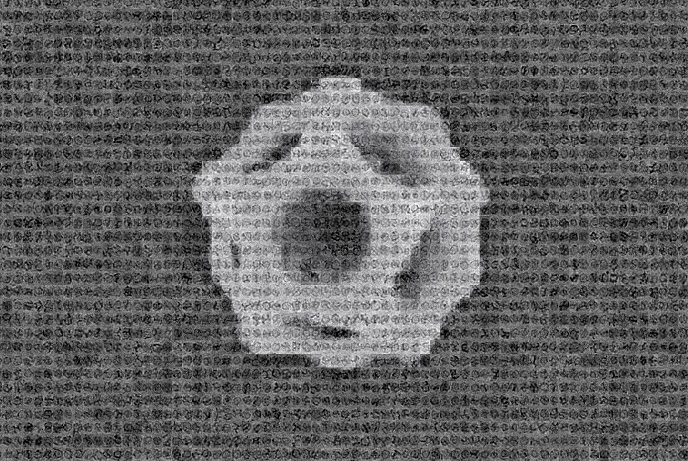
One of the most remarkable project findings was that silica nanoparticles formed a nanocage structure with well-defined dodecahedral symmetry under special conditions. “Studies have shown that tiny silica particles can easily and safely pass through the body and be excreted in the urine, rather than getting stuck in the liver. Perhaps such silica nanocages could be used as containers for anticancer drugs or as vehicles that carry diagnostic molecules,” notes Aubert. The scientists used a relatively simple technique to create this polyhedral structure. For a template, they immersed soap molecules in an aqueous solution. The molecules formed tiny balls called micelles(opens in new window). Then, they added a silica precursor into the mix. Gradually the negatively charged silica clusters formed by the precursor were attracted to the positively charged micelle template surface, building up a cage-type structure. To the surprise of scientists, this process happened without any intervention – the nanoparticles self-assembled around the micelles. “From a fundamental perspective, nanocages offer a unique opportunity to investigate the early stages of the growth mechanisms of ordered mesoporous materials. Observing the self-assembly of nano building blocks into 2D superlattices of silica cages is an additional step in this direction,” adds Aubert. To get a glimpse of these elusive nanocages, scientists used cryogenic electron microscopy(opens in new window). After analysing all different structure orientations by using machine learning algorithms, they reconstructed a perfect dodecahedron.
3D printing sponge-like nanoparticles
Porous nanoparticles have enhanced surface areas that render them highly useful for ‘guest-host’ applications, such as biosensing. They also have better control of the release of therapeutic molecules. By functionalising the silica nanocage with photoresponsive ligands, NanoIntra scientists succeeded in printing mesoporous materials with high surface area and arbitrary shapes. “By placing ligand functionalised silica cages at the desired places of the final printed mesoporous material, we can build highly efficient bioanalytical platforms,” concludes Aubert.