Functional blood vessels revolutionise human 3D tissue culture
Over the years, different animal models have provided invaluable biomedical insight into key pathways and human diseases. However, extrapolating results from animals to drug discovery has proved challenging. Human, in vitro, 3D tissue derivatives such as organoids have received great attention as a means to model the biology of human organs. Technological advances allow the derivation and culture of 3D tissues from stem cells. Accumulating evidence points to their potential for studying human development, but the lack of functional blood vessels significantly limits the recapitulation of the physiology of the native organ.
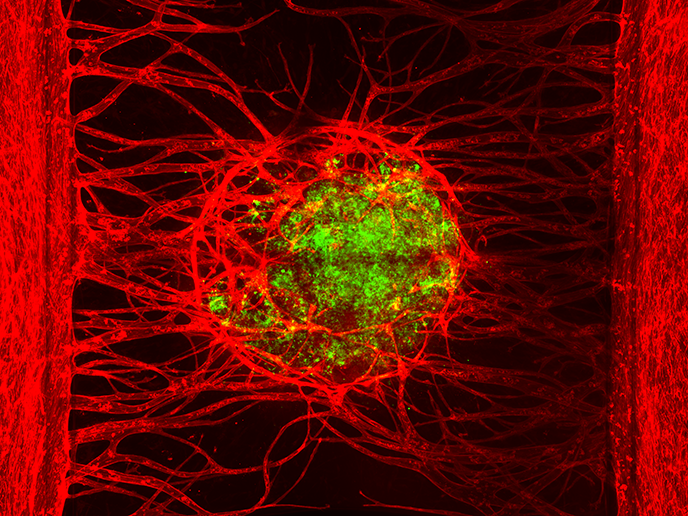
Microfluidics-based platform supporting vascularised tissue
The EU-funded OrganoPlate Graft project sought to address this bottleneck by developing a high-throughput, in vitro culture system for vascularised tissue. The final product of the OrganoPlate Graft project, the OrganoReady Vascular Bed, is a platform of 64 microfluidic chips embedded in a regular, 384-well microtitre plate. The microfluidic chips encompass an extracellular matrix (ECM) gel that facilitates new vessel formation upon administration of a cocktail of angiogenic factors. This essentially creates a vascular bed which can support the grafting of 3D tissues placed on top. Importantly, the microfluidic network in the OrganoReady Vascular Bed offers continuous perfusion of media in a way that mimics natural blood flow. This enables the exchange of nutrients, oxygen and metabolites. “Our product provides researchers throughout the world with the opportunity to add functional blood vessels to their 3D tissues,” emphasises Henriëtte Lanz, project coordinator and director of model development at MIMETAS(opens in new window). Upon connection of the tissues to the vessel sprouts, compounds can be added to the tissue through the vascular network. This allows the study of potential anti-cancer drugs on grafted tumour tissue in an approach reminiscent of patient-derived xenograft animal studies.
Support of hepatic organoids
Organ-on-a-chip technology offers great promise in modelling human liver in vitro; it can be exploited for understanding the process of regeneration as well as for drug discovery purposes. OrganoPlate Graft researchers grafted hepatic organoids and spheroids on the OrganoReady Vascular Bed and established a perfusable vascular network(opens in new window). The 3D liver tissues retained hepatic function throughout the co-culture period and were enriched with microvascular structures. The setup even allowed modelling of veno-occlusive disease(opens in new window) upon exposure to the immunosuppressive drug azathioprine.
Better, ethically produced drugs – without animal experimentation
The OrganoPlate Graft project took on the great unmet need for better models of human biology, required both to speed up basic research and to develop novel, much-needed therapeutic solutions. Implementation of the OrganoReady Vascular Bed in biomedical research is expected to dramatically reduce the need for animal experiments. Moreover, the use of 3D human tissues will support the development of better, more ethically produced drugs. MIMETAS will expand its technology in patient-specific, cancer organoid models for drug discovery purposes with recent funding(opens in new window) from the Dutch National Growth Fund.