Nanosensors for studying neuronal activity
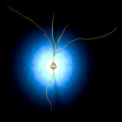
Cell membranes separate internal and external fluids creating electrical and chemical gradients that drive signalling processes. One of the major neurotransmitters, or chemicals responsible for transmitting nervous system information between cells, is glutamate. Binding of glutamate to cell membranes can induce changes in membrane voltage (potential) indicating neuronal activity. Researchers supported by funding of the ‘Voltage-sensitive plasmon-resonant nanoparticles, novel nanotransducers of neuronal activity’ (VSNS) project set out to develop voltage-sensitive nanotransducers (VSNs) for long-term monitoring of neuronal membrane potential to overcome difficulties inherent in using traditional voltage-sensitive dyes. The technologies could be key in developing treatments for neurodegenerative diseases such as Alzheimer’s. Plasmon-resonant nanoparticles (NPs) are metallic NPs that scatter light with remarkable efficiency due to collective resonance (oscillation) of the metal’s conduction electrons. A range of electrically tuneable Plasmon-resonant nanoparticles/nanorods (NP/NRs) acting as voltage nanosensors were developed together with protocols for membrane binding. In addition, the team developed a method for binding and sensing the neurotransmitter glutamate as an indicator of neuronal activity. Single NP spectroscopy enabled insight into fundamental processes linking changes in membrane-bound NP plasmon resonance (NPPR) with measurements of membrane potential. Using simultaneous control of neuronal membrane potential and optical imaging, the researchers studied VSNs bound to membranes in cultured neurons and in cortical tissue slices. Finally, a setup for dark-field microscopy complemented by thermal lensing microscopy (TLM) should be particularly useful for studying changes in intensity, wavelength and phase of scattered light from neuron-bound NP/NRs in preparations such as tissue slices that produce significant scatter. Results of the VSNS project have great potential for use in studying neurodegenerative disease processes and also open the door to a number of new applications in biosensing and optoelectronics.