Novel reconstruction techniques for studying the brain
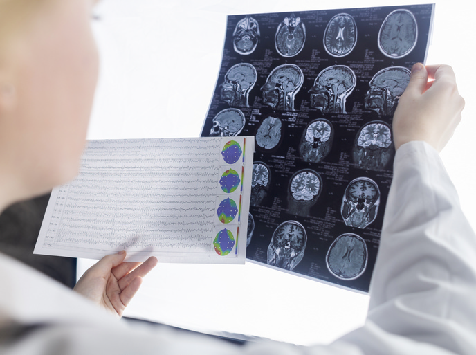
Monitoring of brain activity is instrumental in cognitive neuroscience but also in clinical diagnosis of various brain disorders. In epilepsy, detection of the brain area undergoing seizure is performed by measuring neural activity through implanted electrodes using the intracranial electroencephalography (iEEG) method. However, the invasiveness of this technique causes severe discomfort to epileptic patients. To overcome this, the EU-funded ‘Examining oscillatory dynamics with magnetoencephalography and intracranial electroencephalography’ (Oscillatory Dynamics) project examined the possibility of developing non-invasive methods to monitor brain activity in epilepsy. The primary project goal was to resolve the neural activity of deep brain structures, such as the amygdala, hippocampus, brainstem and cerebellum. Scientists compared the non-invasive technique magnetoencephalography (MEG) with iEEG to map brain activity by recording the electric currents in the brain. The data produced was combined with time-frequency analysis to produce five-dimensional maps of brain activity. Simultaneous MEG and iEEG data was acquired from epileptic patients in order to assess the ability of these two modalities to resolve the activity of deep brain structures. A MEG/EEG analysis toolbox (Nutmeg) was subsequently developed for reconstructing the spatiotemporal dynamics of neural activations and linking them with magnetic resonance (MRI) images. The software was made publicly available at http://nutmeg.berkeley.edu(opens in new window) online. The software was applied and tested on auditory brainstem responses and auditory discrimination following appropriate stimulation. The success of MEG (and possibly EEG) as a tool for examining both normal and pathological hippocampus makes scientists confident that deep brain activity will be monitored more efficiently and less invasively in the future.