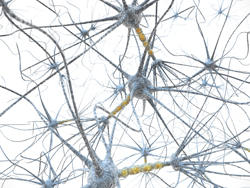
Neuron-level memory formation
During learning, it is believed that synaptic plasticity occurs when presynaptic cells repeatedly and persistently stimulate postsynaptic neurons. This facilitates memory formation and learning through the generation of new synaptic contacts (e.g. dendritic spines on a postsynaptic neuron) or the strengthening of existing contacts. However, researchers are unable to specifically link the memory trace of a behavioural or sensory task to new spine formation. The EU-funded 'Visualizing the structural synaptic memory trace: Presynaptic partners of newly formed spines' (HEBBIANNEWSPINES)(opens in new window) project worked on functionally identifying the presynaptic partners of newly formed spine synapses to determine the neural substrates of memory formation. Researchers employed two-photon imaging systems and optogenetic stimulation methods for their in vitro and in vivo studies. Genetically encoded calcium ion (Ca2+) indicators with dual colour markers were developed to study individual neurons' structure and function during single synapse activation. Ca2+ indicators were used as both dendritic spines and presynaptic boutons experience Ca2+ influx during synaptic activity. For in vitro studies, scientists developed a robust all-optical long-term synaptic potentiation (LTP) paradigm using channelrhodopsin-2 (ChR2) for the selective activation of presynaptic boutons. LTP is the development of a long-lasting increase in signal transmission between two neurons due to repeated stimulation. Study outcomes revealed the formation of new persistent spines over a five-hour period on application of all-optical LTP. Project members had previously demonstrated the formation and stabilisation of new spines during monocular deprivation (MD). MD involves the closure of one eye during presentation of visual stimuli. Researchers successfully developed a combinatorial viral approach for sparse viral transfection of selected subsets of neurons. Imaging the properties of individual spines and large cell populations is now possible. Using the MD paradigm, they can differentiate between signal representation for the open and closed eye and relate structure to function at the synapse level. The tools developed during this project will make it possible to determine if long-term memory formation occurs via spine formation. Understanding the dynamics involved during learning and memory can aid in the rehabilitation of individuals with impaired learning or memory.


