Novel brain tumour imaging
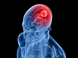
Phosphorous magnetic resonance spectroscopic imaging (31P MRSI) has been gaining attention as an alternative to the well-established MRI. It provides valuable in vivo information about the energy state, pH and metabolism of an area of interest, but resolution is limited and signal-to-noise ratios (SNRs) are low. With advances in high-field (three Tesla (3T)) scanners and multi-channel radio frequency receiving coils, numerous applications are on the horizon. EU-funded scientists set out to develop metrics to assess the aggressiveness of brain tumours with work on the project 'Phosphorus MR spectroscopic imaging of brain tumours at 3T' (31P_SPECTRA_3T). They focused on the spectral peaks produced by the metabolites of interest. The team compared time-domain and frequency-domain analyses of clinical scans for accurate quantification of 31P MRSI data of the human brain at 3T. The low SNRs make this a difficult task. Peak ratio estimates of the two programmes evaluated (Advanced Method for Accurate, Robust and Efficient Spectral fitting (AMARES) and open-source Spectroscopic Imaging, VIsualization and Computing (SIVIC)) were very similar, although AMARES performed better for noisy spectra. Having assessed measurement and quantification techniques, the team turned to application, namely analysing the spatial heterogeneity and characteristics of brain tumours using 31P MRSI at 3T. Scientists compared data from 3 healthy volunteers and 11 patients, all of whom provided legal informed consent. Despite the small sample size, results supported the ability of two techniques (support vector machine and logistic regression) to classify and discriminate brain tumours from normal tissue. Logistic regression resulted in higher sensitivity, specificity and accuracy. Finally, researchers processed the spectra from healthy volunteers and patients using AMARES followed by linear regressions to fit voxel intensities with a given metabolite ratio. They focused on the ratios from the previous assessment that were shown to vary between healthy subjects and patients. 31P_SPECTRA_3T has contributed to greater use of 31P MRSI through enhanced understanding and metrics. This in turn will improve the quality of diagnosis and treatment planning as the databases of healthy and diseased tissues expands.