Modelling the mammalian skull
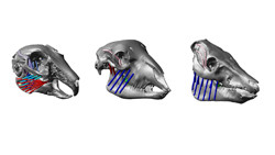
During evolution, mammals have adapted to very different diets. These feeding adaptations are reflected by the diversity in mammalian skull and tooth morphology. However, the relationship between chewing forces and the shape and internal morphology of the jaw bones is not clear, but could help us reconstruct the feeding behaviour of extinct species and better understand the resorption of alveolar bone that is a common cause of tooth loss in the elderly. In this context, the EU-funded 'Examination of alveolar and trabecular morphology and how it relates to masticatory forces' (EAT) project examined the internal jaw morphology of different mammals to understand the link between bone morphology and masticatory forces. The consortium used high-resolution micro-computed tomography imaging to analyse the jaw morphology of different mammalian species including humans, and applied new methods to analyse the trabecular network and thickness of surrounding cortical bone. They also employed magnetic resonance imaging scanning and dynamic computer modelling to generate detailed musculoskeletal models of the whole skull. These models were used to simulate masticatory function and thus make accurate predictions of tooth loading during mastication. Further efforts were devoted to understand orthodontic tooth movement and the role of the periodontal ligament (PDL) fibres in transferring the load from the teeth to the surrounding alveolar bone of the tooth socket. PDL is the fibrous connective tissue that fills the space between the tooth root and the alveolar bone. The scientists showed that by modelling the fibrous structure of the PDL, the alveolar bone is loaded in a way that is not predicted by current hypotheses about the mechanics of orthodontic tooth movement. Taken together, the EAT observations provide important insight into the functional adaptations of the masticatory apparatus in relation to feeding habits and have implications for orthodontic treatments. The generated biomechanical animal models could be further applied in research into animal health and help to reduce the need for in vivo animal models.