Magnetic technology detects breast cancer
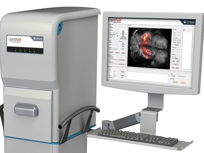
MRI is highly promising when it comes to detecting cancerous breast tissue, but a major issue is a high number of false positives. This not only causes needless patient anxiety, they also have to undergo costly but unnecessary interventions. The advantage of using MRI is that it is non-invasive with no potentially harmful ionising radiations as magnetic fields are used for image production. The EU-funded CADE4BMRI (Improved detection and characterisation of breast cancer using multimodal magnetic resonance imaging and novel computer-aided detection/evaluation (CADe) techniques) study was initiated to resolve issues with specificity and enhance the clinical utility of MRI-based tools for breast cancer diagnosis. CADE4BMRI worked on integrating data from several MRI techniques such as dynamic contrast-enhanced MRI and diffusion weighted imaging via computerised image analysis. The aim was to improve detection and characterisation of breast lesions using parameters like blood perfusion, morphology and tissue microstructure. Project members developed several algorithms for identifying, quantitatively characterising and classifying suspicious lesions in imaging data from breast MRI examinations. These include methods for thresholding as well as supervoxel-based algorithms. Research outcomes were published in a peer-reviewed journal as well as conference papers, and also resulted in a PhD thesis and an MSc thesis. Project members successfully developed a computerised tool with improved sensitivity and specificity for early breast cancer detection. These algorithms are of great interest to developers of MRI computer-aided design systems. Clinical implementation will improve management of breast cancer patients and reduce the deaths resulting from late detection of cancerous tissue.


