New reconstruction methods for parametric imaging
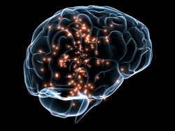
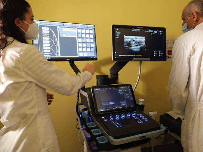
Multimodal imaging is well established in routine clinical practice, especially in the field of nuclear medicine. Compared with computational imaging, magnetic resonance imaging (MRI) offers better contrast amongst soft tissues, minimum radiation exposure to the patient and simultaneous data acquisition. Parametric positron emission tomography (PET) and MRI provide useful insight into a number of neurological disorders such as Alzheimer's disease and aid in characterising brain tumours. Within DYNAMIC PET/MR (Dynamic PET/MR - New reconstruction methods for parametric imaging), researchers improved methods for studying dynamic PET at millisecond range using MRI information. Kinetic modelling of physiological parameters requires an input function (IF) and time-activity curves (TACs). The IF refers to the time course of the activity delivered to the tissue, while TACs of each voxel refer to the variation in activity over time. Using a maximum a posteriori probability reconstruction method in conjunction with anatomical partial volume correction methods, researchers obtained an IF and TACs with less noise and partial volume effects. Using information from MRI, the team developed a new image-derived IF that improved IF estimation in the early frames, which have higher noise and more partial volume effects. DYNAMIC PET/MR methods offer great advantages to medical imaging. Improved image quality through the new reconstruction methods can help reduce a patient's dose. Instead of arterial cannulation or even venous blood sampling, image-derived IF is an attractive non-invasive alternative that makes a patient's therapy more comfortable.