An improved treatment for cardiac arrhythmia
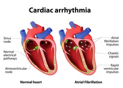
Cardiac arrhythmias are treated or even cured using ablation with a radio-frequency (RF) catheter. However, this approach requires direct contact with the endocardium, thereby limiting the depth of the tissue injury that can be treated. In addition, there is no imaging modality for real-time monitoring of the treatment and for accurate assessment of the lesion. The objective of the EU-funded HIFUSWI-INTRACARDIAC (High-intensity focused ultrasound intracardiac ablation with real-time monitoring using shear-wave imaging) project was to construct an ultrasound-based intracardiac system that could perform ablation in combination with real-time imaging. Towards this goal, the team combined high-intensity focused ultrasound and shear wave imaging (SWI). This allowed them to monitor the size and temperature of the ablation in real time in ex vivo heart samples. The idea was to be able to pinpoint the region to ablate with accuracy alongside a temperature map and close monitoring of the amount of energy to be sent to the tissue. Researchers generated shear waves by the intracardiac transducer at depths of up to 15 mm. They validated and optimised the system in an animal model by monitoring the growth and temperature of cardiac lesions. They also measured the variation of cardiac stiffness over the cardiac cycle and after RF ablation. Importantly, the consortium demonstrated the feasibility of performing real-time RF ablation monitoring in patients using SWI. Overall, the HIFUSWI-INTRACARDIAC system demonstrated the capacity to ablate the cardiac tissue at any location in the thickness of the myocardium while simultaneously assessing the size and temperature of the lesion. This is expected to accelerate procedures, reduce procedure-associated risks and provide a new approach to assess the success of the ablation.