Brain function in an altered oxygen environment

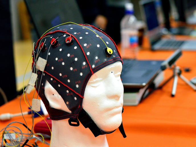
Maintaining O2 concentration at an appropriate level is of outmost importance for the survival of a living organism. Both low O2 (hypoxia) and high O2 levels (hyperoxia) have a deleterious effect on cells. To investigate the outcome of hypoxia and hyperoxia on neuronal function, scientists of the EU-funded BRAINCIROXCON (Functional and structural changes of brain circuitry in altered oxygen conditions) project set out to identify neuron types that are highly sensitive to altered O2 concentration. Using neuroanatomical tools and electrophysiological methods, they mapped the damaged brain areas and identified the damaged inhibitory and excitatory neuronal populations. The most prominent characteristic was cytoskeletal damage -as indicated by the TUNEL assay and staining for apoptosis markers- that was detected mainly in the hippocampus. Intriguingly, even a few hours of mild hyperoxia (30 %) were sufficient to cause neuronal damage. In vivo neuronal damage was validated using magnetic resonance imaging (MRI). Neuronal network activity was measured by electroencephalography (EEG) with multichannel electrodes in the hippocampus under different O2 conditions (normoxic, hypoxic and hyperoxic). Preliminary observations suggested significant changes in network oscillation in hyperoxic and hypoxic conditions as well as increased activity of unidentified inhibitory neurons. To delineate the firing pattern of neurons in response to different O2 levels, researchers employed a single cell method for recording the electrical activity, neuroanatomical and neurochemical characteristics of single cells. No neuronal damage was observed in the prefrontal cortex and analysis efforts concentrated on the hippocampus. Molecular analysis of the damaged neurons is expected to determine the underlying mechanisms governing the role of O2 levels on the observed phenotype. Collectively, the BRAINCIROXCON project provided invaluable knowledge on the impact of O2 levels in the brain. The generated information is expected to aid in understanding the neuronal mechanisms underlying neurodegeneration, and define key regulation steps and direct therapy strategies. Long-term this will improve healthcare and the quality of life of the patients.