Long overdue genetic ID for glial cells
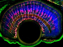
Glial cells, once thought of as the ‘glue’ of the nervous system, are now recognised as having a crucial function in development and function of the brain. Recent research suggests that they may contribute to various neurological diseases such as schizophrenia, autism and even pain. Glial cell populations are very heterogeneous and it is important to distinguish between these when investigating physiology and their precise role in the nervous system. For example, one type of glial cell, an astrocyte develops on injury within the nervous system and subsequently promotes the death of neurons. Despite their importance in physiology and function of the nervous system, little is known about the way in which glia are positioned or shaped to perform their critical support functions. Now, researchers with the EU-funded project Glial Patterning have carried out an in-depth morphological, genetic and transcriptomic analyses of differentiation of a particular type of glial cell, MG cells. Gene identification and activation during the course of developmental cascades “We wanted to investigate the morphological development of MG cells in the retina of zebrafish. Knowledge of this process would serve as a basis for understanding the steps in the post-mitotic differentiation of glia in general,” explains project coordinator Prof. William Harris. Moreover, the results could be constructed to build an experimental paradigm for dissecting the pathways involved in the morphogenesis of these complex cells. The Glial Patterning team was headed up by the postdoc researcher, Dr Mark Charlton-Perkins. Technologies used included clustered regularly interspaced short palindromic repeats, commonly known as CRISPRs, to specifically change genes in the pathways and link their functions with the developing phenotypes, if any. Researchers also identified core candidate genes that have been conserved through evolution. To sort the mixture of phenotypes and correlate them with appropriate genes, the researchers used an efficient fluorescence-activated cell sorting (FACS) method. As Dr Charlton-Perkins points out, “We were lucky at the time to have a good way to FACS sort MG cells at several stages.” Next steps for MG research “Results of the investigations look extremely promising on all fronts,” says Prof. Harris. The scientists identified genes expressed at each developmental stage and a range of interesting phenotypes that correlate with each of these. “We are now writing up the results.” The plan for future research of MG cells is very extensive. Prof. Harris points out, “Each of the genes that have been discovered will need more thorough investigation. In particular, questions of autonomy and mechanisms of action will need to be addressed.” Glial Patterning research results have the potential to form a solid knowledge platform for future research into the functions of MG cells in health as compared to disease phenotypes. A detailed molecular picture of all the genetic and transcriptomic stages in the development of MG cells could help identify target molecules for new therapies.