

German scientists capture structure of tuberculosis membrane
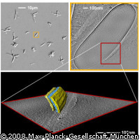
German researchers have obtained the first ever 3-D images of a double-membrane surrounding the mycobacterium responsible for tuberculosis. Their findings have ended a long debate on the structure of the mycobacterial outer membrane and are expected to offer new opportunities for drug development. TB is a deadly infectious disease that most commonly attacks the lungs, but can also affect the central nervous system, the lymphatic system, the circulatory system, the genitourinary system, bones, joints and even the skin. It is estimated that each year around ten million people fall ill with TB, while 4,000 people die every day from the disease. The disease is caused by a bacterium called Mycobacterium tuberculosis, and it is difficult to treat because of the mycolic acids (long fatty acids found in the cell walls of the mycobacterium). The structure of these acids lends the organism increased resistance to chemical damage and dehydration, and prevents antibiotics from penetrating the cells. Up until now, it was believed the mycolic acids formed a closed layer around the cell or were part of a considerably thick and asymmetrical membrane. Now, scientists from Max Planck Institute of Biochemistry in Martinsried, Germany, have proved that architecture of the outer cell wall is structured somewhat differently. Rather than an asymmetrical membrane, the researchers discovered that the cell wall was composed of a distinct lipid bilayer. Using an electron microscope, they investigated the cell structure of Mycobacterium smegmatis and Mycobacterium bovis BCG, a close relative of the tuberculosis bacterium. Then, by means of cryo-electron tomography, the scientists were able to obtain 3-D images of the bilayer structure from intact cells. The method requires projection data from different angles of a shock-frozen cell (-190 °C). In order to avoid radiation damage, the cell must only be exposed to the electron beam for a limited period of time. With the findings of their research, the scientists at Martinsried will now conduct a more detailed study of the mycobacterial outer membrane. The outcome is expected to pave the way for the development of new chemotherapeutic drugs. 'After all, the drugs must pass through cell wall as effectively as possible, and a better understanding of the mycobacterial cell envelope will certainly be helpful,' says Dr Harald Engelhardt, who led the research.
Countries
Germany