

Scientists develop innovative technology to fight cancer
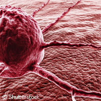
EU-funded researchers continue the good fight against cancer. A Dutch team has succeeded in boosting chemotherapy dosage to tumours with the possibility of easing harmful side effects by combining magnetic resonance imaging (MRI) and ultrasound. Their research is funded in part by the SONODRUGS ('Image-controlled ultrasound-induced drug delivery') project, which is backed with almost EUR 10.8 million under the Nanosciences, Nanotechnologies, Materials and new Production Technologies (NMP) Theme of the EU's Seventh Framework Programme (FP7). Eindhoven University of Technology collaborated with Philips Electronics to create sophisticated technology with the potential to help patients in need. The team performed a demonstration of this technique for the first time ever in a preclinical, proof-of-concept study. Around 50% of patients diagnosed with cancer undergo chemotherapy to treat their illness. Chemotherapy drugs flow through the bloodstream, damaging rapidly dividing cells like cancer cells. But in their quest to fight disease, chemotherapy drugs know no boundaries and end up attacking normal, healthy dividing cells found in various parts of the body including bone marrow, hair and mucosae. Patients then face a number of side effects, such as neutropaenia, a lower white blood cell count, and anaemia, a lower red blood cell count, as well as bleeding. Some researchers say chemotherapy is complex in that the drugs are spread everywhere in the body and are not absorbed evenly; some regions fail to get the dosage they need. Headed by Eindhoven's Holger Grüll, the team created a delivery method to tackle this problem. An MRI scan would initially be performed on cancer patients; this would help doctors localise the site and size of the tumour. Liposomes, small temperature-sensitive particles containing an MRI contrast agent and a chemotherapy drug, are injected into the patients and move through the bloodstream until they reach their target, the tumour. The contrast agent moves along with the drug allowing it to follow the path of the drug inside the body and localise when it reaches the tumour and surrounding tissue. At this point a high-intensity focused ultrasound (HIFU) beam is used to burst the liposomes and release the drug at the site of the tumour. For the purposes of their study, the researchers assessed the uptake of the anti-cancer agent doxorubicin in rats. They found that uptake was between two and five times higher in induced tumours versus regular chemotherapy delivery. 'In some tumours, you have a centre part that is no longer supplied with blood, and so the chemotherapy cannot reach that part and cannot kill it,' said Matthew Harris from Philips Research. The team said the prototype system is based on a Sonalleve 3-Tesla MRI-HIFU scanner, which is used to treat uterine fibroids - painful benign tumours formed from the muscular layer in a woman's uterus. The MRI scanner combined with high-intensity ultrasounds pinpoints and removes the fibroids via ablation, the removal of unwanted cells. This proof of concept appeared in the Journal of Controlled Release in February 2011.For more information, please visit: Eindhoven University of Technology:http://w3.tue.nl/en/SONODRUGS:http://www.sonodrugs.eu/
Countries
Netherlands