Taking precise photos of proteins - is filming them just round the corner?
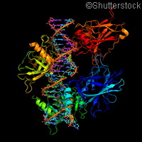
An international team of researchers has found a more effective way of imaging proteins, something that could soon lead to filming how they work at the molecular level. The team, from Germany, Sweden and the United States, built on previous work led by one of the study authors, Professor Richard Neutze from the University of Gothenburg, who was one of the first in the world to image proteins using very short and intensive X-ray pulses. The better scientists can map out the structure of proteins and how they behave in cells, the closer they can get to unlocking the cure to major diseases such as cancer and malaria. This new study, published in the journal Nature Methods, tests out the method on a new type of protein, and the results bode well for future experiments. The team looked at a membrane protein from a type of bacterium that lives off sunlight. It is important to investigate membrane proteins, as they transport substances through the cell membrane and thus take care of communication with the cell's surroundings and other cells. Lead study author Linda Johansson, also from the University of Gothenburg, comments: 'We've managed to create a model of how this protein looks. The next step is to make films where we can look at the various functions of the protein, for example, how it moves during photosynthesis. To put it simply, we've developed a new method of creating incredibly small protein crystals. We've also shown that it's possible to use very small crystals to determine a membrane protein structure.' There are two major challenges when it comes to imaging proteins: the first is to create the right-sized protein crystals, and the second is to irradiate them in such a way that they do not disintegrate. Although Sweden has a facility for synchrotron-generated X-ray radiation at Lund University, this type of technology is not light-intensive enough, and therefore requires large protein crystals that take several years to produce. To overcome this hurdle, Professor Neutze decided to try and image small protein samples using free-electron lasers that emit intensive X-ray radiation in extremely short pulses - shorter than the time it takes light to travel the width of a human hair. Luckily, the research partners from California could provide a facility that fitted the bill. 'Producing small protein crystals is easier and takes less time, so this method is much faster,' continues Linda Johansson. 'We hope that it'll become the standard over the next few years. X-ray free-electron laser facilities are currently under construction in Switzerland, Japan and Germany.' A key discovery was that far fewer images are needed to map the protein than previously believed. Using a free-electron laser it is possible to produce around 60 images a second, which meant that the team had over 365 000 images at its disposal. However, just 265 images were needed to create a three-dimensional model of the protein.For more information, please visit:University of Gothenburg:http://www.gu.se
Countries
Germany, Sweden, United States


