Fast imaging of the heart by ultrasound
Ultrasound is a very attractive means of assessing the morphology and the blood flow in the heart chambers as well as for monitoring the motion of the cardiac walls. Importantly, it operates in real-time, offers good temporal resolution and doesn’t use ionising radiation. However, current solutions offer insufficient time resolution to evaluate all details of the heart dynamics, requiring fast echocardiography.
Building a 3D ultrasound scanner for heart imaging
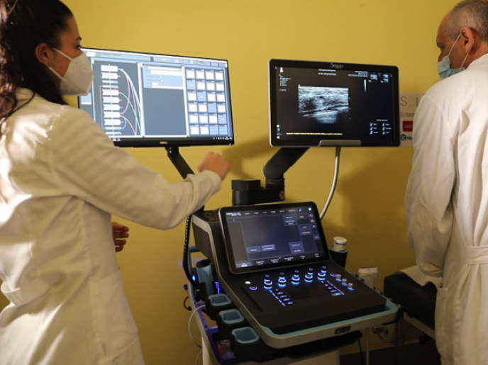
Undertaken with the support of the Marie Skłodowska-Curie (MSC programme, the aim of the ACOUSTIC project was to overcome existing technical limitations in both 2D and 3D heart ultrasound and develop an innovative system that offers very high temporal and spatial resolution. “Our goal was to reliably and reproducibly measure the very fast motions of the heart muscle during the cardiac cycle,” explains the MSC fellow Alessandro Ramalli. The ACOUSTIC system is based on the ULA-OP 256(opens in new window) research echograph which offers great versatility and programmability. Researchers implemented non-conventional fast imaging techniques and processing approaches for imaging blood flow and motion of the heart muscle with unprecedented detail. Moreover, they integrated a fast imaging mode that reconstructs three imaging planes simultaneously. Improvement of 3D heart ultrasound relies on a prototype ultrasound probe, whose layout was inspired by the seeds in a sunflower, that allows 3D imaging while maintaining low cost. In principle, this probe can be connected to any commercial scanner without the need for additional expensive devices.
Significance and future perspectives
Preliminary data on patients demonstrate the capacity of the ACOUSTIC system to achieve high spatial and temporal resolution of wide fields of view in both 2D and 3D echocardiography. Important features are retention of good image quality and real-time operation, significantly enhancing the diagnostic power of echocardiography. “Contrary to other fast imaging solutions that rely on the offline processing of acquired echocardiography data, the ACOUSTIC system gives real-time feedback of the results to the physician,” emphasises Ramalli. The system allows the analysis of rapid cardiac events offering a better knowledge of regional contractile function with direct clinical implications such as for the improved diagnosis of conduction disorders. It also allows for the robust assessment of haemodynamics of the whole heart and not just a narrow region of interest. From an industrial perspective, the project has demonstrated the possibility of delivering echocardiography at high temporal resolution with relatively low-cost hardware already available in commercial scanners. This has the power to expedite the implementation of the ACOUSTIC features in clinical practice worldwide. Ongoing efforts focus on technical optimisation by developing novel techniques for the improvement of cardiovascular image quality and the post-processing capacity of the system. Recruitment of more volunteers and patients in project trials will further validate the clinical utility of the ACOUSTIC ultrasound system for cardiovascular disease diagnosis. Considering the increase in the ageing European population and the growing socioeconomic burden of cardiovascular diseases, advanced imaging modalities endow diagnostic and prognostic power to the cardiac ultrasound examination.


