The tiny gland with a big impact on breathing

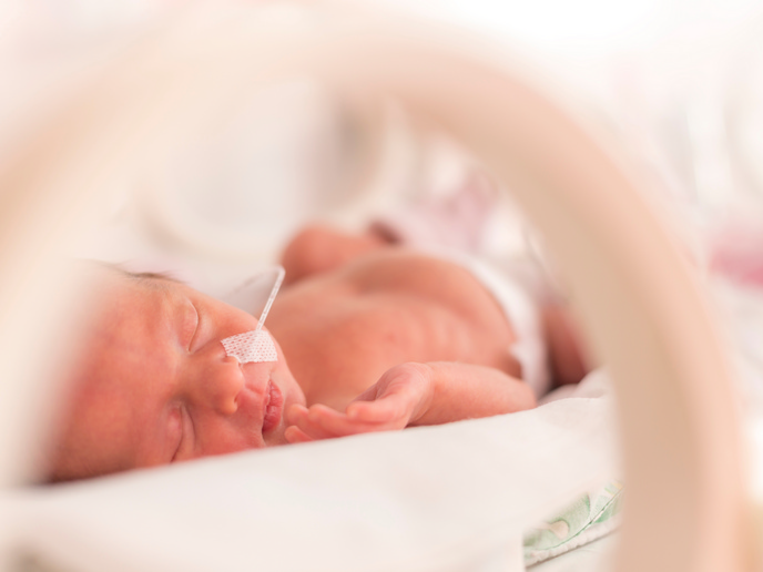
Diseases of the respiratory system kill more than 340 000 people every year in the EU, around 7.5 % of all deaths(opens in new window). The EU-funded OxygenSensing(opens in new window) project sought to shed light on the molecular mechanisms by which the body senses oxygen levels in the blood in order to respond to them. Humans have evolved adaptations to combat low oxygen levels in the blood – a condition known as hypoxia. These can be rapid reactions to acute oxygen shortages, such as hyperventilating during physical exertion, or longer lasting adaptations, such as increasing the number of red blood cells in response to chronically low blood oxygen resulting from lung disease or life at high altitude. Acute changes in blood oxygen are detected by the carotid body, a small, pea-sized gland found adjacent to the arteries at both sides in the neck. “The carotid body is connected directly to the respiratory centre in the brain, and when oxygen decreases, it increases the breathing rate and heart output,” explains project coordinator, José López-Barneo. His laboratory at the University of Seville(opens in new window), in Spain, has been working on the carotid body for 35 years. Yet, says López-Barneo, there was a basic unanswered question: how the carotid body cells detect changes in blood oxygen.
Chemical sensor
His team looked at glomus cells, chemoreceptors that are found in the carotid body. Years ago they identified potassium channels that closed during hypoxia, triggering a signal to be sent to the brain. Yet how these potassium channels are modulated by oxygen has remained elusive. Through their work on the OxygenSensing project, López-Barneo and his team were able to identify mitochondria in these cells as the key oxygen sensor. Usually, mitochondria are able to function normally even in extremely low oxygen environments, far beyond what is experienced in physiological hypoxia. But López-Barneo’s team discovered(opens in new window) that in the glomus cells of the carotid body, the mitochondria use an unusual variant of the enzyme cytochrome C oxidase. “We found glomus cells’ affinity for oxygen is much lower than normal, and the electron transport chain changes within the physiological range of oxygen availability,” he says, meaning a modest decrease in blood oxygen disrupts mitochondrial activity in these cells. This generates signals that close the potassium channels, leading to the secretion of transmitters that activate sensory nerve fibres. These fibres tell the brain to increase breathing and heart output.
New treatments
“This is the basic discovery, now we have a solid model available, and the predictions of this model can be tested experimentally,” adds López-Barneo. “Our ongoing work confirms that the model is experimentally robust.” The research may go some way to explaining a curiosity of COVID-19, where patients develop acute hypoxia but do not react physically to it, so-called ‘silent hypoxia’. López-Barneo hypothesises that the virus may disable chemoreceptors in the carotid body in the same way it does in the nose and mouth, causing a loss of taste and smell. The clinical applications of the findings are widespread. López-Barneo says the research will help support the development of powerful respiratory stimulants based on activation of an oxygen sensor in the carotid body. This would be hugely beneficial in anaesthesia and in treating opioid overdose.