A new atlas of the brain reveals its connections in unprecedented detail
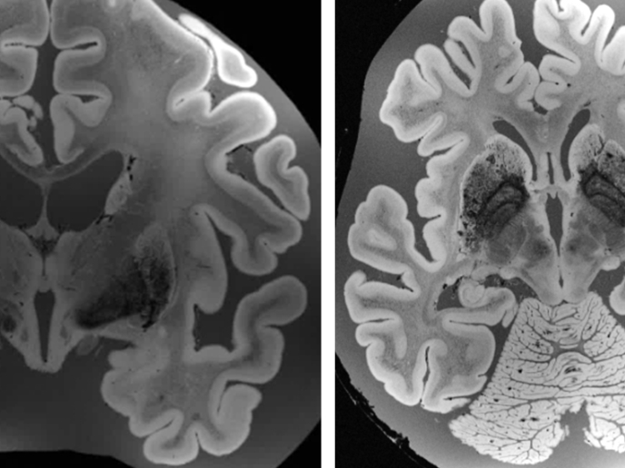
The human brain contains an estimated 86 billion neurons(opens in new window) and an equal number of support cells, arranged in complex networks and circuits, the architecture of which is still largely unknown. The EU-funded MULTICONNECT project sought to shed light on the structure of the brain through the use of pioneering imaging techniques. In the past, the most common method for studying the anatomy of the human brain was to cut thin sections around 100 microns thick and examine these under a microscope. However, doing this destroys and therefore obscures the 3D structures in the organ. “If you take the cortex, you have to look at it over a sufficiently large field of view to have a perspective of its complicated 3D structure,” explains project coordinator Alard Roebroeck. “We want to look at the brain in a multiscale and multimodal way, to have a perspective measured in centimetres, but at the same time a resolution of single cells.”
High power
To achieve this, Roebroeck and his team, from the Multiscale Imaging of Brain Connectivity section in the Department of Cognitive Neuroscience at Maastricht University(opens in new window), took large pieces of neural tissue from cadavers, including entire brains, and imaged them with an ultra-high field strength MRI scanner. “The MRI scanners in hospitals typically operate at 1.5-3 tesla and have a resolution around 1 mm isotropic,” says Roebroeck. “We used up to 9.4 tesla, using custom-built radio-frequency coils and longer imaging times, to image the whole brain at 100 microns and below.” This data was then combined with images captured through light sheet fluorescence microscopy(opens in new window). This technique illuminates samples with a flat sheet of laser light, allowing the interior of the tissue to be imaged in thin optical sections without disrupting its 3D structure and at very high imaging speed. To maximise the preservation of 3D structures, Roebroeck used large sections of brain tissue up to 5 mm thick, first making them transparent by removing unwanted material such as lipids. In order to handle samples this large – measuring 8 cm square and larger – a custom light sheet microscope had to be designed and built. Roebroeck and his team were able to visualise fine details, such as the individual layers, columns and cells of the cortical sheet, itself just 4 mm thick. “We are first to image this at 75 micron resolution across the whole brain,” he adds.
Data processing
The project generated huge amounts of data, with data for a single sample containing up to 10 terabytes of information. “There are always concomitant challenges with advances in imaging, you need new ways to adapt and process and deal with data that is a thousand times what you’re used to,” notes Roebroeck. The work was supported by the European Research Council(opens in new window). The imaging techniques have already attracted interest for their potential in other fields, such as tumour analysis in pathology, and implant placement in brain stimulation. The new atlas of the brain structure will be made available to other researchers. A key question for Roebroeck is how the microstructure of the brain defines the kind of computations it can carry out. “We’re starting to change the view of computation in the brain. Now we know better what constraints there are, and the types of circuitry that can be formed in those cells,” he says.