Mapping gene expression in two colours

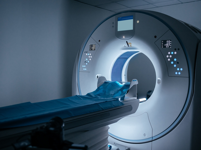
MRI is an important tool used in clinical practice and medical research. Since its invention in the 1970s, MRI technology has helped doctors and medical scientists detect, monitor and track a variety of medical conditions, its performance increasing with every advancement made over the last few decades. A new method developed by researchers from Israel and the United States could improve the effectiveness of MRI even more. With support from the EU-funded GeneREFORM and AutoCAb projects, the research team have found a way to use MRI to track two different genes, in two colours.
Why this achievement is important
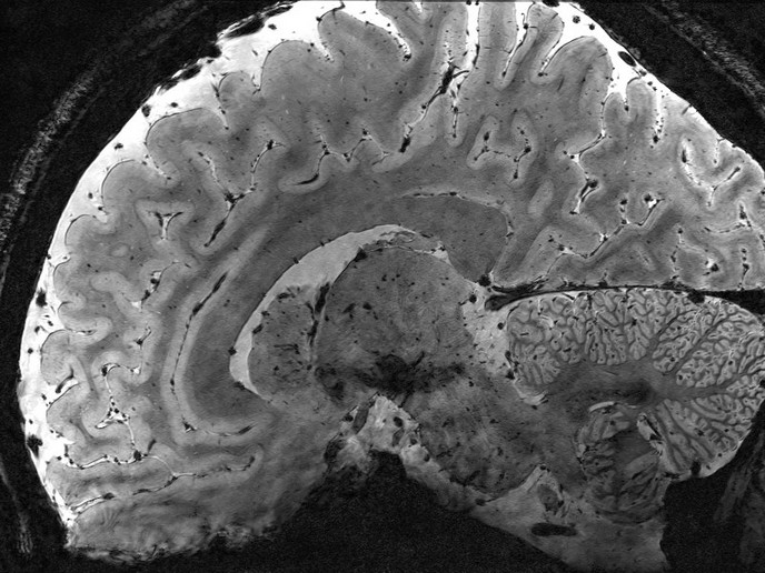
Being able to map out real-time gene expression in our body’s cells using contrasting colours would provide invaluable insight into currently invisible biological processes. The glowing multicolour proteins that scientists currently use in fluorescence microscopy to track gene expression are not much help when it comes to observing what goes on in the deep tissues of the body. While MRI today enables scientists to observe such deep-seated processes, the grayscale images produced do not provide specific information about gene expression. The researchers’ new method is now making this possible. “MRI may one day be used to peer deep into the body over an extended period of time, to see what goes on in tissues without the need to remove them for study under a microscope,” remarks Dr Amnon Bar-Shir of GeneREFORM and AutoCAb project host Weizmann Institute of Science, Israel, in a news item(opens in new window) posted on ‘Phys.org’. “Our method provides a major step in that direction,” continues Dr Bar-Shir, corresponding author of the relevant study(opens in new window) published in the journal ‘Nature Biotechnology’. The team genetically engineered two groups of highly active reporter genes, each group expressing one of two specially designed enzymes. They also designed two novel molecular probes that are MRI-detectable, unlike fluorescent probes whose signals can be blocked by thick tissues. These reporter probes are injected into the bloodstream and accumulate exclusively in the reporter genes expressing the specially designed enzymes. This makes it possible to track the reporter gene expressions through the signal that the probes emit in response to different MRI frequencies, with each probe showing up in a different colour on the MRI map. “Gene expression lets us know what each cell is doing,” states the study’s lead author Dr Hyla Allouche-Arnon, also from the Weizmann Institute of Science. “Thanks to our method, MRI may now be applied by researchers in various fields to track the activity of all kinds of processes, for example, those involving different types of brain or immune cells.” The researchers tested their method on live mice using extremely powerful MRI equipment with a magnet of about 15 tesla. The scans detected the two probes’ signals and revealed the precise positions of the cells expressing the engineered proteins, showing them in green and pink. The method devised with support from the GeneREFORM (Genetically Encoded Multicolor Reporter Systems For Multiplexed MRI) and AutoCAb (Automated computational design of site-targeted repertoires of camelid antibodies) projects could be further developed to simultaneously map more than two genes in different colours. In the future, if adapted for use in humans, it could be used to non-invasively monitor important processes such as the progress of cancer treatments. For more information, please see: GeneREFORM project(opens in new window) AutoCAb project(opens in new window)