A high-performance PET scanner for integration into current MRI systems
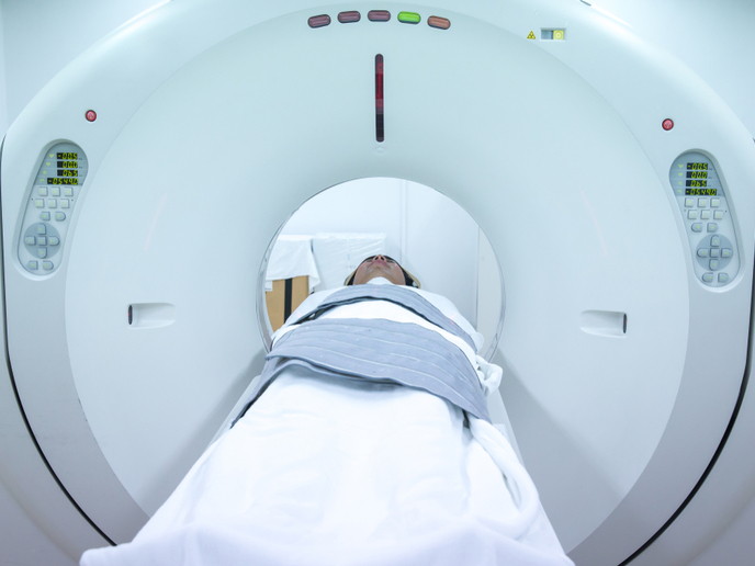
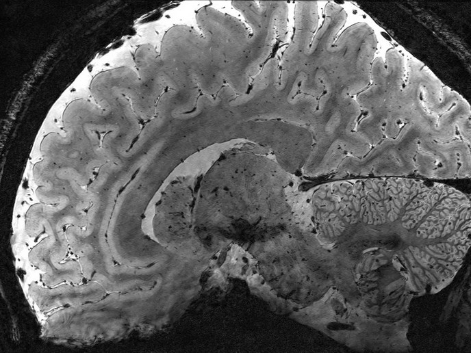
Besides its high resolution and efficiency, the main advantages of MINDVIEW’s (Multimodal Imaging of Neurological Disorders) new PET imager are its significantly reduced cost, its size and the fact that it allows for conducting both Positron Emission Tomography (PET) and Magnetic Resonance Imager Radio Frequency (MRI RF) imaging at the same time. “MRI and PET provide complementary information,” explains Prof. Jose Maria Benlloch Baviera, coordinator of the project and director of the CSIC’s Institute for Instrumentation in Molecular Imaging (I3M). “MRI provides high quality and resolution images on the morphology of the soft tissue, which is very useful in localising lesions in the body. On the other hand, PET images provide information on the physiologic processes occurring in different organs and substructures. The two technologies potentiate each other: For instance, MRI information can be used to better position physiologic information from PET within the brain.” The potential for the precise and early diagnosis of mental disorders such as schizophrenia and severe depression is tremendous. Neurotransmitter pathways-specific PET radiopharmaceuticals – such as glutamatergic, serotonergic or dopaminergic – can be imaged, while areas of the brain activated by performing a task are tracked with functional MRI. According to Prof. Benlloch Baviera, this might eventually lead to precise and quantitative diagnosis of mental disorders that would be impossible to obtain with currently available techniques. MINDVIEW’s PET system also stands out through its design. Compared to current devices made of thousands of small crystal pixels coupled to an array of photo-sensors, this one includes only 60 large monolithic crystal blocks coupled in one side to a matrix plane of silicon photo-sensors. “This design has several performance advantages. Since the same amount of light is emitted in all directions, we can determine the depth of gamma ray interactions by measuring the width of the light distribution. In other words, we can find the 3D position of the gamma ray impact. This is a critical feature to avoid image blurring when using scanners that are close to the organ. Besides, light is directly detected by the sensors instead of bouncing back and forth in the pixel. This allows in principle for better energy and timing resolution, which in turn reduces signal noise at the image,” Prof. Benlloch Baviera explains. Such design also brings cost down considerably, as tasks such as cutting, polishing, painting and gluing thousands of pixels back into a single block are avoided. This is a major incentive for hospitals which generally must pay between EUR 4 and 7 million for commercial PET/MRI systems. Smaller clinics that cannot afford a new whole-body PET/MRI system might even be able to finally acquire this powerful technology. Last but not least, the new design allows for a portable system that can easily be removed when it’s not needed for a particular MRI examination. As Prof. Benlloch Baviera points out, this represents a major technical breakthrough since the mutual interference of MRI and PET imaging modalities, in particular when being physically so close to each other, would normally be substantial. “For instance, the high field (3 Tesla) produced by the main magnet of the MRI may completely degrade the performance of the photo-sensors and electronics of the PET scanner. Special photo-sensors based on silicon technology have been developed and non-paramagnetic electronics and connections have been used in order to avoid that effect,” he explains. “Furthermore, innovative shielding of the PET modules and the RF coil have been designed to avoid eddy currents that will distort the high homogeneity of the magnetic field required for a top-quality MRI image.” The device is currently being tested on patients with Alzheimer’s disease (AD).