Advancing quantitative MRI for better patient care
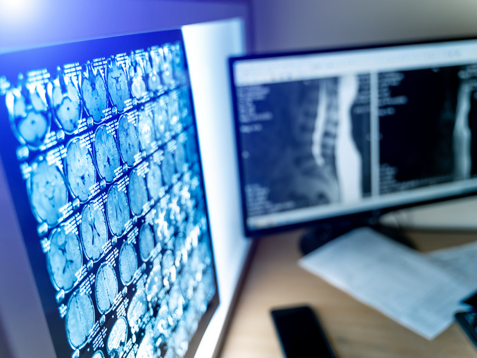
With almost 100 % sensitivity and specificity, for many, magnetic resonance imaging(opens in new window) (MRI) is the gold standard in imaging. This is particularly the case when it comes to looking at soft tissues, such as the brain, with MRIs providing radiologists a much clearer picture of the organ than an X-ray or a computerised tomography (CT) scan. Yet even MRI has its shortcomings. “Although MRIs excel at providing highly detailed images, these images are qualitative,” says Jan Sijbers, a professor at the University of Antwerp(opens in new window). “This means the resulting contrast is highly dependent not only on the underlying tissues, but also on the type of scanner and protocol used to capture the image.” This qualitative nature of MRI images can hinder a radiologist’s ability to perform a quantitative comparison of tissue properties within a scan, between successive scans, and between subjects – all of which can be essential to reaching a proper diagnosis.
Precise measurement of brain features
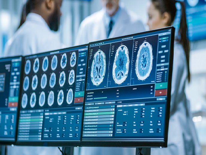
This limitation has led to the development of quantitative MRI (Q-MRI). “In Q-MRI, the contrast is directly related to the underlying biophysical characteristics of the soft tissue, which substantially improves clinical diagnosis,” explains Sijbers. “The problem is that Q-MRIs involve a long scan time, making them uncomfortable for the patient and expensive for the clinic.” With the support of the EU-funded B-Q MINDED project(opens in new window), Sijbers is spearheading an effort to close the gap between qualitative and quantitative MRIs. “Our goal was to transform MRI from a pure imaging modality into a quantitative tool for the precise measurement of brain features.”
Improving patient care
One of the project’s key results was the development and validation of super-resolution reconstruction (SRR) methods. These methods allow a radiologist to reconstruct clear, high-resolution images from a single-frame, low-resolution image. With these methods, project researchers were able to compute high-resolution Q-MRI parameter maps using a set of low-resolution, qualitative MRI images. “The use of SRR methods substantially reduced acquisition time while retaining spatial resolution and signal-to-noise ratio,” adds Sijbers. Researchers also developed methods for data harmonisation and robust estimation of diffusion parameters. Another important achievement was the conducting of successful T1 (longitudinal relaxation time) and T2 (transverse relaxation time) mapping based on recurrent inference machines – an achievement that resulted in a 100-fold reduction in the time needed to infer quantitative maps. “These results will allow radiologists to more easily and objectively reach conclusions about the status of brain disease, which ultimately improves patient care,” notes Sijbers.
Further advancing Q-MRI
According to Sijbers, this Marie Skłodowska-Curie Actions(opens in new window) supported project’s success is the direct result of the 15 young, bright early-stage researchers (ESRs) involved in the work. “It was a real privilege to be a part of this dynamic team and watch as they worked together to develop and effectively implement quantitative MRI methods in preclinical practice,” he says. “They made this the most interesting and exciting project of my career.” These researchers will now take the experience and knowledge they gained during the project and apply it towards further advancing Q-MRI. At the same time, Sijbers will soon start working with a new generation of ESRs on a project that looks to incorporate artificial intelligence into Q-MRI methods.