New imaging system helps to optimise stem cell therapy for arthritis

Arthritis is a crippling condition and the most prevalent disease in the world. Osteoarthritis, caused by the gradual wearing of cartilage in joints over time, affects around 10% of the global population. There are roughly 70 million patients in Europe alone. Yet there is still no effective cure. Most treatments focus on alleviating symptoms and do not restore form or function. Stem cells present one promising therapeutic avenue, offering a unique opportunity to regenerate injured cartilage. This therapy is hampered though, by an inability to detect successful engraftment in real time using conventional imaging techniques. “The promise of stem cell treatments is restrained by the lack of knowledge of where the cells go and what they do in humans,” explains Martin Leahy(opens in new window), chair of Applied Physics at the University of Galway.
Restrictions in current technologies
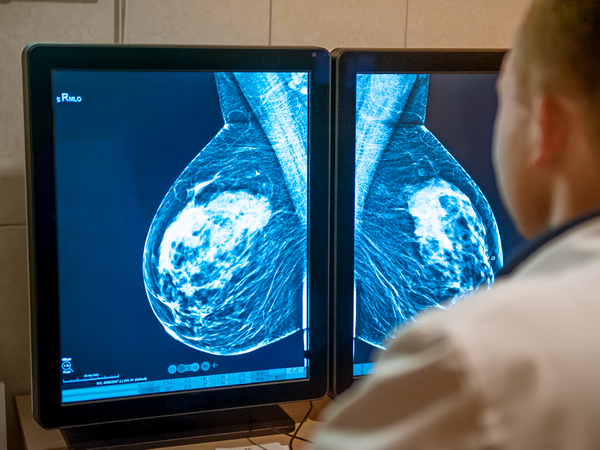
Current imaging technologies cannot detect items smaller than 1/200th of the tissue depth, and stem cells have low intrinsic contrast, which makes them difficult to see. “This restricts progress on their healing properties and safety,” says Leahy. Leahy coordinated the EU-funded STARSTEM(opens in new window) project, which proposed an innovative new imaging technique able to capture human stem cells at clinically relevant levels. STARSTEM introduced the use of gold nanostars, which absorb 10 times more light than conventional dyes and help to pinpoint stem cells. “We were able to see mesenchymal stem cells(opens in new window) (MSC) and extracellular vesicles(opens in new window) (EV) deep in sheep knees, the best model of human knee osteoarthritis, for several months after injection,” Leahy notes. MSCs are cells isolated from a mix comprising bone marrow, and EVs are tiny vesicles within MSCs.
Imaging with nanotechnology
STARSTEM aimed to harness and improve the properties offered by photoacoustic imaging, a non-invasive system that uses lasers to construct images of tissue – and other substances – under the skin. Nanomaterials possess certain properties which enable them to enhance this kind of imaging system. The STARSTEM researchers internalised gold nanostars into stem cells, which enhance contrast and improve deep-tissue imaging. “The STARSTEM nanostars were optimised to provide the greatest energy deposition and hence photoacoustic contrast of any particle,” Leahy explains. “The energy deposited in the nanostars causes the surrounding cells and tissue to vibrate more than with any other particle, so they become more easily visible with the photoacoustic system,” he adds. The team optimised every part of the imaging process, allowing them to detect the smallest number of MSCs and EVs at clinically relevant depths. Through a series of trials, they were able to find the optimal size and shape of gold nanoparticles to improve the imaging contrast. These advances allowed the team to maximise the uptake of stem cells while ensuring no toxic effects.
A pathway to regenerative medicine
The advances could help scientists better understand how stem cells work, and aid in the transition from preventative to regenerative medicine. This branch of medicine aims to create methods to repair or replace lost cells. Both MSCs and EVs are thought to trigger healing and support tissue repair within the body. The STARSTEM team is now aiming to test their gold nanostars in clinical trials.