Deep brain activity in Parkinson’s disease
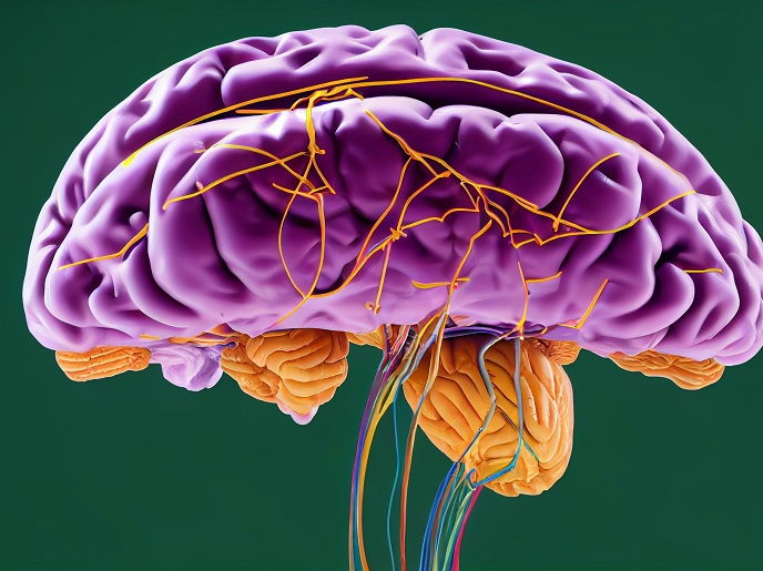
Parkinson’s disease (PD) is associated with a loss of dopamine producing neurons in the substantia nigra(opens in new window), a part of the brain that helps control movement. PD patients experience motor symptoms such as tremors, rigidity, as well as gait disturbances. Emerging evidence indicates the implication of another two deep brain structures, the mesencephalic locomotor region(opens in new window) (MLR) and the subthalamic nucleus(opens in new window) (STN), in gait disturbances and falls in PD patients. The STN is a key basal ganglia nucleus involved in the regulation of movement, and pathological activity in this structure is associated with motor symptoms as well as abnormal gait and postural instability in PD patients. At the same time, abnormal activity in the MLR can result in difficulties with initiating and maintaining gait.
Insight into deep brain activity in Parkinson’s disease
The key objective of the LINKERS project was to better characterise the abnormalities of deep brain activity that could explain some of the gait disorders and/or falls in PD patients. The research was undertaken with the support of the Marie Skłodowska-Curie Actions(opens in new window) (MSCA) programme and focused on the STN and MLR brain structures. Researchers recorded the activity of these deep brain structures in PD patients who underwent surgery for deep brain stimulation(opens in new window) (DBS), a procedure that involves targeted implantation of electrodes in a specific part of the brain and the delivery of electrical pulses. The electrodes in these patients were implanted in the STN or in the MLR. The PD patients were asked to walk on an instrumented gait platform that allowed the collection of neurophysiological and biomechanical data. Leg muscle activity was also recorded to study the relationship between brain activity and muscle activity. “We were able to demonstrate a temporal and spatial relationship between gait initiation and activity in the STN or MLR,” outlines MSCA research fellow Yannick Mullie. A reduction in specific brain activity in the STN was associated with the ability of patients to take large steps or walk fast. Moreover, patients who experienced motor block or freezing of gait demonstrated an increase in brain activity of a different frequency. Higher brain activity was also evident in the MLR of these patients.
Neural activity during freezing of gait
Freezing of gait is a challenging symptom to manage and significantly impacts the quality of life of people with PD. It is characterised by a sudden and temporary inability to start or continue walking, despite the intention to move. Scientists aimed to understand the brain activity modifications that occur during and before the occurrence of this blockage. To achieve this, they recorded the activity of the STN and MLR regions during episodes of freezing of gait. Although no clear neuronal pattern was evident, an increase in specific brain activity in the STN of specific patients occurred at the beginning of freezing. Collectively, LINKERS results indicate the presence of a functional STN-MLR network for locomotion and postural control in humans. This will serve as the foundation for the development of DBS patterns tailored to patient needs and in accordance with specific walking and/or balance disorders. According to Mullie: “The next step is to correlate leg muscle activity with neural activity to realise an adaptive stimulation that improves the quality of life of PD patients.”