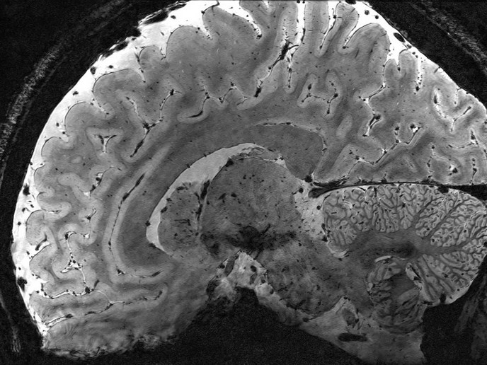
Hybrid fMRI-PET scans offer insights into the ageing brain
Functional magnetic resonance imaging(opens in new window) (fMRI) reflects the increase of blood supply to active regions of the brain, providing the oxygen and nutrients needed by the neurons. Comparing the fMRI signals of individuals across different age groups or the same individuals, over time provides insights into the architecture and workings of the human brain. This has shed light onto normal and pathological age-related brain changes and of individual differences, helping detect and characterise neurodegenerative disorders, such as Alzheimer’s disease. “However, some common fMRI patterns, such as the deactivation of certain brain areas during cognitive control and increased activation of prefrontal areas in ageing, have led to questions of interpretation,” notes Anna Rieckmann(opens in new window), project coordinator of the SIMULTAN project, which was funded by the European Research Council(opens in new window). SIMULTAN used positron emission tomography(opens in new window) (PET) scanning to complement fMRI signals, as it can pick up additional signals connected with age-related changes. “We showed that hybrid fMRI-PET scanners can visualise complementary aspects of brain function simultaneously,” says Rieckmann.
Supply and demand in the ageing brain
When neurons are active, they require more oxygen and nutrients (metabolic demand), so the blood vessels in that region dilate, increasing blood flow and oxygen supply (haemodynamic contributions). This leads to an oversupply of oxygenated haemoglobin in the area, visible to fMRI imaging. The problem is that the many processes which link the firing of neurons with increased fMRI signals are complex and not fully understood. “For example: Are the differences in fMRI signals of older adults, compared to younger adults, reflecting differences in neuronal activity or the blood flow and oxygen supplies of older vascular systems?” asks Rieckmann.
The best of both worlds
To identify typical changes in brain activation, SIMULTAN scanned the brains of 30 young adults (20-30) and 40 seniors (65+) with a hybrid fMRI-PET scanner, while they performed a memory task. PET is a molecular imaging method that monitors brain glucose metabolism, triggered primarily by synaptic activity and unaffected by changes in blood flow. The team were specifically interested in the fMRI signals from the areas of the brain that could help compensate for an ageing brain, while the PET signals reflected metabolic demand in these regions. Increased fMRI signals in older adults were not found to be associated with higher metabolic demands, suggesting differences in fMRI activity are not a result of neurons working harder but are rather due to an ageing vascular system. Furthermore, when older adults performed a task as well as younger adults, their neural metabolic demands were also equal. “Maintaining cognition into old age is likely a reflection of brain health rather than other brain regions compensating,” remarks Rieckmann.
Longer lasting brain health
It is often assumed that ageing inexorably heralds cognitive decline. But many older adults still maintain cognitive fitness, with SIMULTAN finding 75-year-olds still able to perform cognitive tasks like 40-year-olds. If we can pinpoint the factors that maintain cognitive well-being, we can identify targeted interventions. “With fMRI alone it is difficult to identify intervention targets because signals are not specific to a neurotransmitter, pathway or protein, and while PET gives that specificity, it isn’t great at portraying overall brain function. But combined imaging could lead to interventions that delay neurological illness,” concludes Rieckmann. SIMULTAN’s approach will now be applied to altered brain function after physical exercise, to ascertain whether changed fMRI signals reflect the effects of training on blood flow or on neuronal firing.