Zooming in on microbial life
Every animal is considered an ecosystem in its own right, colonised by microbiota in organs such as the skin and the gut. Together with the host, these microbial communities make up what scientists call the holobiome. Understanding the interactions amongst these commensal microorganisms and with the host is central for animal health, welfare and production. Traditional methods of studying host-microbiota interactions focus on microbe identification but overlook a significant parameter: the precise location of microbiota within the host, their proximity to nutrients and competitors. All these can have profound consequences on how microbes behave and interact. Therefore, there is a need to unveil the spatial context of bacteria and biomolecules in tissues.
Integration of omics technologies
To understand how microbes interact with each other and with the host, the EU-funded 3D-omics(opens in new window) project set out to use a combination of omics(opens in new window) approaches, tools that analyse genes, proteins, and chemicals in the body. By mapping this information in 3D, the team wanted to see not just what is happening in the gut, but exactly where it’s happening. According to project coordinator Antton Alberdi: “By integrating multiple omics data from host and microbes we can move beyond simply cataloguing which organisms are present and begin to understand what each of these organisms is doing and how they interact.” By looking at which genes are switched on, which proteins are synthesised, and which small molecules are present, scientists can obtain information on how microbes and host influence each other. When combined, these layers of information provide a clearer picture of how these relationships impact animal health.
Analysing microbes in 3D
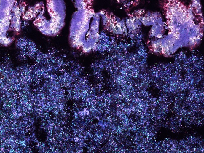
Each type of omics analysis requires its own sample preparation and processing protocols, making it hard to combine results. Also, many reference databases often lack essential information on the function of microbial genes. To complicate matters even further hosts and microbes are differentially regulated, and microbial metabolites can affect parts of the body far from their site of production. To solve these issues, the consortium pioneered a new method called microscale spatial metagenomics (MSSM). Unlike conventional omics, which require large amounts of material and ignore 3D structure, MSSM identifies which microbes, genes and functions are present in 25-micrometre-wide grids. This enables researchers to know exactly which strains associate with probiotic species, or how pathogens like Salmonella position themselves near host tissues.
Turning microbial maps into meaningful insight
The 3D-omics team has applied the MSSM platform to improve livestock production, bringing to light how specific strains interact with native microbes in the gut and where they colonise. This 3D information enables researchers to pinpoint how microbes cluster around specific nutrients such as fibres, added in feed to support metabolism and immune function. MSSM provides information on where fibre degradation happens and which microbes work together, enabling the design of targeted fibre blends and supplements that support beneficial gut ecosystems more effectively. “Our micro‐scale microbiome analysis technology can be applied to any complex microbial ecosystem,” highlights Alberdi. The team is now extending its research to human health to predict which microbial communities will flourish under specific diets or treatments. In the future, the 3D-omics approach could help tailor probiotic therapies to individual patients or livestock breeds by accounting for the specific spatial structures of their gut microbiota.