Smart microscopy sheds light on how cells organise
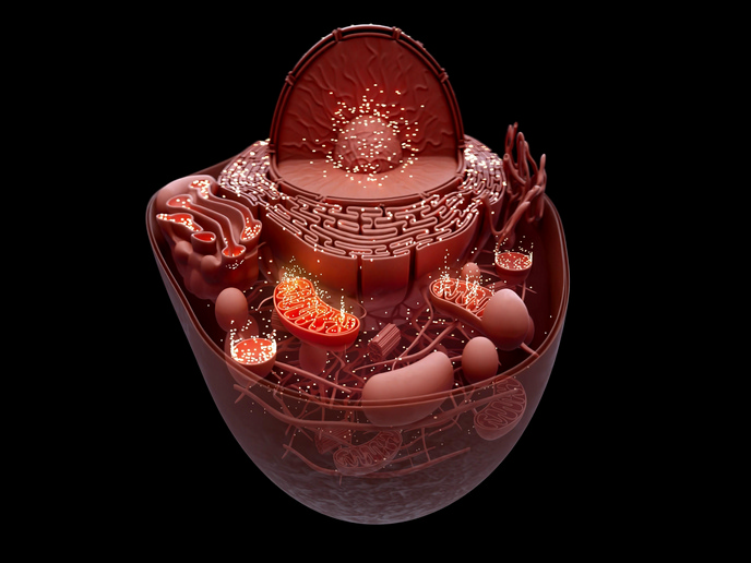
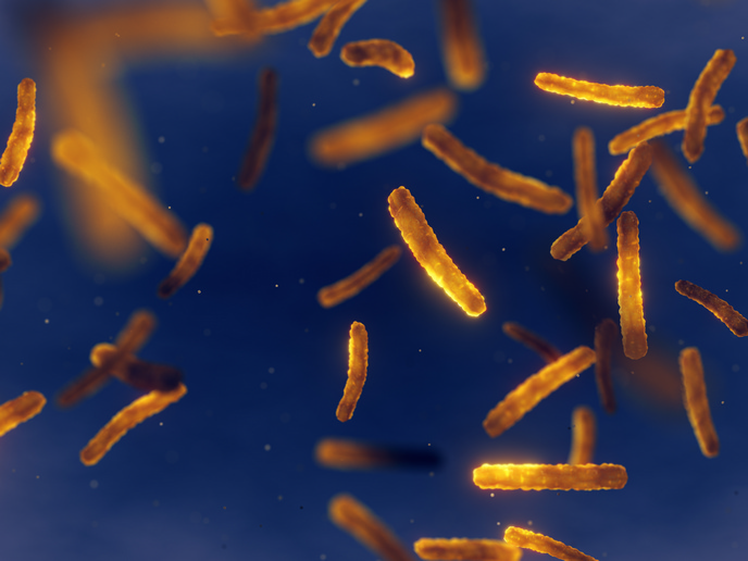
The insides of eukaryotic cells contain structures called organelles(opens in new window), specialists in a variety of vital functions such as generating energy (mitochondria) and storing genetic material (nucleus). Cells are so crowded that the molecules inside them, like proteins and nucleic acids, regularly bump into each other, inspiring the Piko project, which was funded by the European Research Council(opens in new window). As Suliana Manley(opens in new window), project coordinator explains: “We wanted to understand how crowding might affect dynamic processes, as too much road traffic creates bottlenecks, slowing everything down.” Piko was especially interested in parallels between mitochondria and bacteria; mitochondria having evolved from bacteria and with each carrying their own DNA. The team investigated how crowding impacts the organisation of DNA as it is replicated (copied) and segregated (split apart), in both bacteria and mitochondria. “While simpler than human cells, bacteria can better survive harsh conditions, typically existing in a quiescent or low-activity state, to endure periods of starvation,” says Manley. “The complex behaviour of the bacterial cytoplasm is important for their survival, but how it affects processes like DNA organisation is poorly understood.”
Smart microscopy offers ‘gentler’ biological discovery
To unravel the organisation and dynamics of the interior of bacteria and mitochondria in living cells, the team faced imaging challenges. Firstly, with only a small number of molecules involved, detecting telltale signals is tricky. Secondly, the processes are transient, occurring only over a brief period within the cell cycle. Thirdly, the features of interest are often too small to detect using classical light microscopy. “Cells and mitochondria are sensitive to light, plus we needed to avoid disrupting their function while we watched,” adds Manley. The project adapted different forms of microscopy which required less light, advancing three areas: smart microscopy, mass photometry super-resolution, and multi-parameter mapping of objects with optical nanoscopy (MOON). While the last two remain at the proof-of-concept stage, Piko significantly developed smart microscopy, offering gentler forms of microscopy for biological discovery. The team also trained a neural network to recognise certain mitochondrial shapes which triggered the microscope. “We monitored cellular dynamics using a gentle white light, activating fluorescence only when the neural network detects a mitochondrial constriction. Or we used light-intensive super-resolution microscopy to monitor activity over large time intervals, triggering short time interval images when constriction begins,” notes Manley. Simulations helped identify patterns by comparing bacterial cytoplasm or mitochondrial matrix organisation against random models. In bacteria, how DNA is organised was found to have direct implications for replisome(opens in new window) dynamics – the multi-protein machine that replicates DNA. In mitochondria, geometric and molecular patterns in division were discovered that determine distinct organelle fates. For example, dividing in the middle leads to proliferation, while dividing at the tip leads to degradation. The team also discovered how mitochondria distribute their DNA, via a transformation of the normally tubular organelle into a string of pearls shape.
Wider health research benefits
With researchers increasingly using smart microscopy for a wide range of applications, such as studying infection, the benefits of Piko’s advances could prove game-changing. And with mitochondria playing many important roles in human health and disease, researchers are already using project findings for insights into a broad range of pathologies, including neurodegeneration and cancer. In the meantime: “There are still mysteries about how some of the patterns we discovered emerge, and how they interact with each other, which I would love to solve,” says Manley. “We also want to better understand how mitochondria coordinate with partner organelles, or each other, within cells to decide which functions to carry out, and when.”