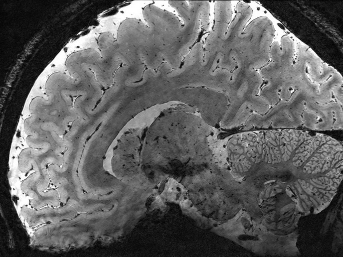
Magnetic resonance studies of the brain

Ten European Magnetic Resonance (MR) centres have collaborated on this research project in order to develop, using three different MR techniques, standard quality assurance and clinical protocols and to apply these new techniques to both diseased and healthy human brains. The central aim being the identification of pathophysiological factors important for diagnosis and treatment monitoring. Magnetic resonance spectroscopic imaging (MRSI), water diffusion imaging and perfusion imaging were the three techniques applied in the project. With spectroscopic imaging a single slice metabolic imaging pulse sequence was developed in order to obtain quantitative metabolite concentrations. The centres participating in the project had to implement the pulse sequence in their respective MR scanners; which has lead to the choice of a multi echo sequence with a single spatial phase encoding step per cycle. In vivo studies were performed on rats. In order to obtain a standard, measurements of the developed, MRSI protocol were compared with measurements of T1 and T2 of water and metabolites, and with calculations of metabolite concentrations. Measurements were performed at four different centres on six normal volunteers each. The research showed that MRSI results are appropriate for purposes of comparison and interpretation. For diffusion imaging a multi slice echo-planar spectroscopy sequence was used. Anisotropy effects are eliminated if the values of the anisotropy tensor at three perpendicular distances, i.e. its trace, are obtained while applying two magnetic field values at each one of the directions. Validation studies are still in progress in this area. In invasive perfusion imaging, the assessment of cerebral perfusion and blood volume was facilitated by the use of the contrast agent Gd-DTPA during fast imaging. The contrast agent, a paramagnetic ion (Gd) bound up in a large molecule (DTPA), that enhances the local magnetic fields, was bolus injected and followed through its path with 6 anatomical slices per second. The critical aim of the project, through better understanding of the human brain, its physiology and metabolism, is not only, to ease the economic burden brain diseases impose on health care systems, but above all to ease the emotional burden on sufferers and their families.