A virtual gallery of medical pictures
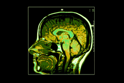
Magnetic resonance imaging (MRI) is a medical imaging technique that uses a magnetic field and radio waves to create high-quality images of the head or body, without the need for X-rays or any other harmful forms of radiation. An MRI scan provides information on brain structure that is essential for early diagnosis of neurodegenerative or brain disorders such as Alzheimer's or Parkinson's disease. The major problem lies in defining what constitutes a 'normal' brain, given the huge variability of brain morphology. In order to model this variability, an enormous number of brain images from patients of all ages, genders and backgrounds are required. Albeit an important tool for such modelling, a European repository of this kind does not currently exist. An investigation was needed to forecast what is required in order to develop a large and shared European multidimensional repository of MRI scans of both 'healthy' brains and those with disorders. This was the objective of the 'Foresight study for the development of an European neuroimage repository' (ENIR) project. ENIR investigated the standardisation topics required and the practical implications of acquiring, processing and storing neuroimages. This was done by means of a coordinated approach to establishing a devoted research infrastructure. The project also identified the standardised procedures for making the best use of existing repositories. In the long term, the ENIR results may pave the way for a pilot prototype of the repository with a restricted number of medical centres. Once operational, the system could be extended to other centres, and expanded with other medical images such as positron emission tomography (PET) scans. It could also be used to aid research into new treatments and, in the long term, benefit patients with neurodegenerative disorders.