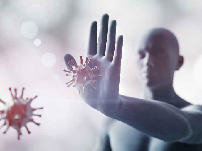
Illuminating cellular and molecular mechanisms in real time
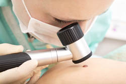
Current imaging modes such as magnetic resonance imaging (MRI), computed tomography (CT) or positron emission tomography (PET) can identify relatively small tumours at the organ or tissue levels. There is however a need for imaging technologies that can provide cellular and molecular information in vivo for cancer prevention, detection and targeted therapy. CARS EXPLORER was a multidisciplinary EU collaboration working towards the development of NLO imaging methodology to detect skin cancer using mouse and human melanoma (skin cancer) models for reference and validation. For this purpose, scientists investigated NLO microscopy techniques such as coherent anti-Stokes Raman scattering (CARS), two photons fluorescence (2PEF), and second harmonic generation (SHG) for sub-cellular visualisation in biological samples. Pulse shaping techniques were used to optimise NLO contrast, incident power, penetration depth and signal propagation in microscopes and endoscopes for deep tissue imaging. Hollow core (HC) photonics crystal fibres (PCFs) were exploited to deliver high-power femtosecond (10−15 second) pulses in the form of solitons in endoscopes. Solitons are self-reinforcing solitary wave packets or pulses that travel at constant speed while maintaining their shape. Researchers were successful in setting up point scanning microscopes that used compact Ti:Sapph lasers and optical parametric oscillators. Customised software was developed for system control and data processing. The stimulated Raman scattering (SRS) method was used to create image contrast in tissue images using non-linear microscopy. This was covered as a News Feature in Nature magazine. An endoscope-microscope system prototype using CARS signal and SRS was built for in vivo and in situ visualisations of samples; patenting is in process. Databases were successfully generated that could characterise and differentiate between normal and cancerous skin and lymphoid tissue using Raman and CARS spectroscopy. Label-free mapping was also done of nucleic acids, proteins and lipids in melanoma skin tissue sections. Tumour expressing mouse models were generated for exploring the efficacy of these techniques in living animals. These novel technologies have demonstrated their potential for early detection and diagnosis of cancer and other diseases by identifying malignant cellular changes in real time.