

Benefits of scientific cooperation under the microscope
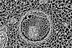
An EU-funded project has brought together some of Europe’s leading electron microscopy facilities in order to provide tailored access to academic and industrial partners. This cross-border collaboration has helped to open up new research avenues that were not available before, and underlined again the strength of European research cooperation. Electron microscopy uses a beam of electrons to create an image of an object. These microscopes are used to investigate the ultrastructure of a wide range of biological and inorganic specimens including microorganisms, cells, large molecules, biopsy samples, metals, and crystals. Industrially, the electron microscope is often used for quality control and failure analysis. Modern electron microscopes produce electron micrographs using specialised digital cameras and frame grabbers to capture the image. While being capable of much higher magnifications and greater resolving power than light microscopes – allowing scientists to see much smaller objects in finer detail – the equipment needed is large and expensive. It often requires specially designed facilities and trained personnel to operate. By pooling resources therefore, ESTEEM2 has been able to provide access to such facilities to a much wider audience. The ESTEEM2 project granted access to applicants via a simple peer review process based on merit and scientific priorities. Users were then supported via a networking programme that sought to address key issues such as specimen preparation, data interpretation and standardisation of methodologies. In addition, a series of schools and workshops were organised to provide training in innovative electron microscopy methods and an online forum established for discussing emerging cutting-edge techniques. For example, a workshop on advanced transmission electron microscopy (TEM) was held in Sweden in July 2015. TEM is powerful tool for research into materials, nanostructures and nano-devices because it can provide information about the structure and chemistry of substances at the atomic level. In addition, electron energy loss spectroscopy (EELS) and other types of spectroscopy have the potential to further expand scientific understanding of structure and properties. Another workshop took place in Cadiz, Spain. This workshop focused in particular on transmission electron microscopy of nanomaterials, highlighting the advances that have been made and the range of nanomaterials that have been examined. Participants were given the chance to discuss their data, ask questions and receive advice on carrying out future experiments. Scientific cooperation facilitated by the project has also led to new discoveries. By coupling a scanning transmission electron microscope with an interferometer – used to measure tiny displacements or surface irregularities – new single-photon emission (SPE) sources were successfully detected. This confirmed the possibility of detecting single-photon emission sources from different materials with a novel experimental setup. The discovery could lead to further searches for new SPEs in different materials, including 2D monolayers. The ESTEEM2 project, due for completion at the end of September 2016, has helped to consolidate Europe’s strategic leadership in electron microscopy, while materials developed through during the project will continue to guide future experiments and promote electron microscopy to the wider research community at large. For further information please visit: ESTEEM2 project website(opens in new window)
Countries
France