Development of new MRI capabilities
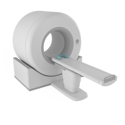
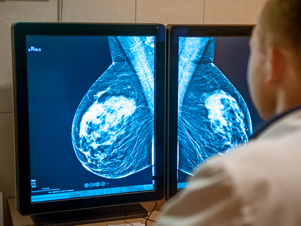
MRI has a wide range of applications in medical diagnosis as it does not use any ionising radiation. The EU-funded Marie Curie 'Magnetic resonance methods development and applications for life sciences' (EUROCANMRI) project aimed to develop new diagnostic capabilities in the areas of cancer, spinal cord and Alzheimer's diseases. Conventionally, MRI images are formed by applying gradients to the main static magnetic field. However, the gradient equipment is expensive, complex, and can induce circular currents in nearby conducting structures, including the patient tissues. Over the initial period of the project the MRI console was designed, successfully installed and tested with a range of phantoms for hydrogen and phosphorus resonances. The project team continued development of software packages for different imaging techniques with particular focus on gradient-free MRI methodology. The results with this approach demonstrated one-dimensional and two-dimensional imaging capabilities and slice selection using a single-channel MRI system. Potential applications include a low-cost process that could be used in a space station setting. Early diagnosis of brain tumour glioma is crucial and the scientists investigated different contrast agents for diagnostic purposes. The study showed that contrast agent the project had developed, anti-IGFBP7 iron oxide single domain antibody, selectively targets abnormal vessels within a mouse glioblastoma. In addition to research and development work, the project team organised a workshop. About 70 MRI scientists from Canada, USA and European countries participated in the event exchanging ideas and knowledge. Project partners presented six talks and two posters illustrating their work.