MRI for Huntington's disease
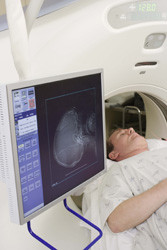
Huntington's disease (HD) is an inherited disorder characterised by progressive degeneration of brain cells, and is caused by a mutation in the gene that encodes the protein huntingtin (htt). This leads to impairment of movement and cognitive disability, and may also reach psychiatric dimensions. Although genetic testing can predict disease onset long before it develops, currently there is no cure. For most efficient disease management and monitoring of preventive treatments, HD biomarkers are urgently required. With this in mind, the EU-funded MULTIMODAL MRI IN HD project set out to develop tools for measuring signs of disease pathology. The idea was to devise methods for evaluating neural change over time, especially before symptoms arise. In this context, scientists developed a magnetic resonance imaging (MRI) protocol and combined it with advanced imaging analysis. They looked at iron distribution in the brain of pre-symptomatic and early-stage HD patients, discovering that, even before symptoms emerge, there is a progressive iron increase and volume loss in basal ganglia. Interestingly, these features were strongly associated with the type of mutation. This led scientists to conclude that iron accumulation may be linked with the observed toxicity underlying HD. Furthermore, HD patients exhibited structural brain impairment in the form of grey matter loss in all the cortical and sub-cortical areas. Brain damage seems to inflict more peripheral nodes first, indicating that rehabilitation may be a viable intervention. Emerging treatments for neurodegenerative diseases come in the shape of iron chelators and antioxidants. The findings of the MULTIMODAL MRI IN HD study not only support the rationale of such modalities, they also underscore the importance of assessing iron levels and tissue integrity. Combined with relaxometry, MRI and grey/white matter measures could prove useful for monitoring the onset and progression of HD.


