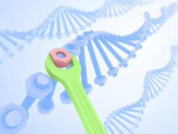
Visualising DNA repair
Cells have evolved to maintain the integrity of their genetic material through sophisticated mechanisms. When DNA gets damaged, for example by irradiation, it undergoes double-strand breaks (DSBs) that get repaired through specialised enzymes. Improper repair of DNA can lead to the formation of cancer. Following damage, DSBs are not the only events that take place but are strongly linked with changes in the chromatin structure of the DSB vicinity. This is believed to facilitate the access of repair proteins to DNA in order to restore genomic integrity. With this in mind, the EU-funded project 'Study of protein dynamics in living cells after DNA damage' (LCS) set out to analyse the dynamics of DNA damage with particular focus on chromatin-related proteins. LCS researchers were interested in comprehending how the process of chromatin opening is coordinated to provide access to the DNA repair machinery. For this purpose, they induced ultraviolet-mediated DNA damage in both mouse and human cells in vitro and followed the kinetics of a number of nuclear proteins by fluorescence imaging. Using adopted bioinformatics, they were able to associate changes in fluorescence with protein localisation. They made the interesting observation that the protein Oct-4 is capable of recognising DNA lesions and that additional transcription factors get recruited in areas of DSBs. Furthermore, the research team investigated how epigenetic modifications influence DNA repair. They discovered that an overall rearrangement of the epigenetic pattern follows DNA damage. Collectively, the findings of the LCS study provide fundamental knowledge on the mechanism of DNA repair after damage. The generated results have a translational impact as they provide enhanced understanding of how cancer develops as well as of the events following treatment by irradiation.


