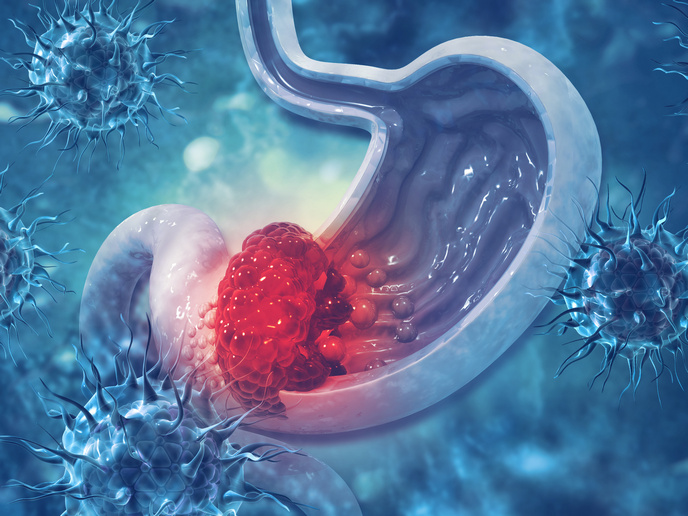
Improving histology classification
Human observation is currently the most reliable way to diagnose and grade cancer from histology slides, yet there is significant inter- and intra-rater variability. That variability is likely to decrease significantly with image processing technology for major impact on patient treatment and prognosis. With EU funding of the project 'Microscopic image processing, analysis, classification and modelling environment' (MIRACLE)(opens in new window), scientists developed a valuable tool to aid experts and enhance the reliability and accuracy of diagnoses and grading. Recognising the invaluable and necessary role of the expert given variability and nuances present in human tissues, the tool is meant to support and not replace expert opinion. Through training and exchange among five universities in Europe and the United States, researchers developed microscopic image processing algorithms and software for images of follicular lymphoma (FL), neuroblastoma and cancer cell line images. Registration with the MIRACLE FL database enables the user to download the software from the project website or upload cell line images for classification, annotation and download. Medical experts can upload their images to support expansion of the available database. The MIRACLE project has received extensive press coverage around the world. Articles are available on the project website. The team published several papers and organised numerous international workshops and special sessions at conferences. The work will culminate in a special issue organised by project scientists in Signal, Image and Video Processing (Springer) on the topic of microscopic image processing. Increasing the accuracy of cancer diagnosis and grading can both reduce underestimations of severity as well as enhance efficiency of treatments. Both of these benefit patients tremendously. Exploiting the power of computing and image processing will now enable experts to do their jobs even better.